Femoral Head Fracture without Dislocation by Low-Energy Trauma in a Young Adult
- Affiliations
-
- 1Department of Orthopedic Surgery, Seoul National University College of Medicine, Seoul, Korea. oskim@snu.ac.kr
- KMID: 1097173
- DOI: http://doi.org/10.4055/cios.2011.3.4.336
Abstract
- We describe the case of a healthy young man with a femoral head fracture by low-energy trauma that occurred without evidence of hip dislocation. While plain radiographs showed no definite fracture or dislocation, computed tomography (CT) and magnetic resonance imaging (MRI) revealed a femoral head fracture with a wedge-shaped cortical depression at the superomedial aspect of the femoral head. Our patient reported feeling that the right hip had been displaced from its joint for a moment. This probably represented subluxation with spontaneous relocation. The characteristic findings and possible mechanisms of this fracture were postulated on the basis of the sequential 3 dimensional-CT and MRI. The clinical results of conservative treatment were better than those of previously reported indentation fractures.
MeSH Terms
Figure
-
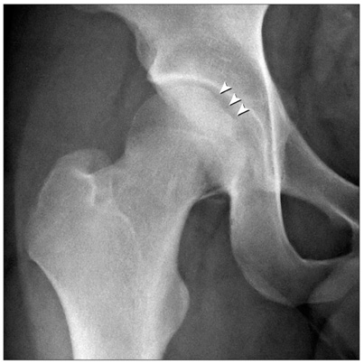
Fig. 1 Initial plain radiograph only showed a cortical defect at the superior aspect of the fovea of the right femoral head.

Fig. 2 Initial computed tomography showed a wedge-shaped cortical defect at the superomedial aspect of the femoral head (asterisk) with fracture extension (arrowhead).

Fig. 3 Computed tomography reconstruction of the isolated right femoral head showed three-dimensional morhpology of the fracture.

Fig. 4 Initial fast spin echo T2-weighted fat suppression image (A) showed joint effusion and a wedge-shaped osteochondral indentation with the fracture line extended to the inferior portion of the femoral head with bone marrow edema. Follow-up magnetic resonance imaging (MRI) (B) revealed femoral head fracture healing with residual reactive bone marrow edema. Final follow-up MRI (C) showed a completely healed femoral head fracture.

Fig. 5 Fast spin echo T2-weighted serial images on axial plane showed changes in fracture healing (A: initial, B: seven months after injury, C: nine months after injury).

Fig. 6 Radiograph at nine months after injury showed mild joint space narrowing and a marginal osteophyte (arrowhead).
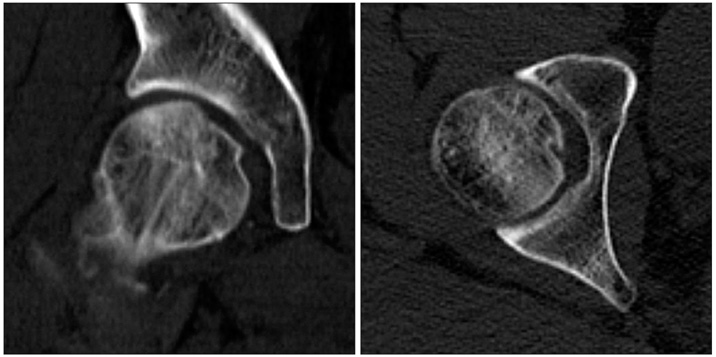
Fig. 7 Nine-month follow-up computed tomography scans showed union of the fracture with minimal flattening at the superior aspect of the femoral head and residual cortical depression.
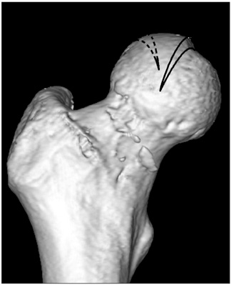
Fig. 8 Solid line represents the direction of the fracture of this patient and dotted line shows the direction of previously reported indentation fractures.
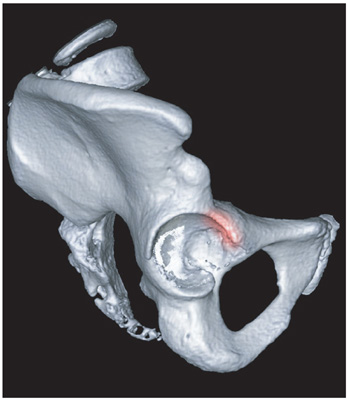
Fig. 9 The anterior edge of the acetabular rim (red zone) is supposed to be causative area against which impingement of the femoral head occurred in this patient.
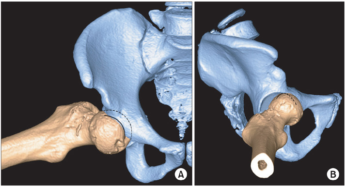
Fig. 10 The fracture of this patient was simulated using three dimensional reconstructed computed tomography images by positioning the femur in abduction, external rotation, and flexion. (A) Solid line represents the fracture line of this patient and dotted line indicates outline of the femoral head. (B) The fracture line is schematically drawn as black line around the femoral head (dotted line indicates the fracture line at hidden area).
Cited by 1 articles
-
Traumatic Femoral Head Fracture without Hip Dislocation - A Case Report -
Ji Wan Kim, Hyun-Wook Chung, Taek-Soo Jeon, Hyung-Nam Shim, Tae-Yeon Yoon, Young Chang Kim
Hip Pelvis. 2012;24(3):256-260. doi: 10.5371/hp.2012.24.3.256.
Reference
-
1. Matsuda DK. A rare fracture, an even rarer treatment: the arthroscopic reduction and internal fixation of an isolated femoral head fracture. Arthroscopy. 2009. 25(4):408–412.
Article2. van der Werken C, Blankensteijn JD. Fracture of the femoral head without dislocation: a case report. Acta Orthop Scand. 1987. 58(2):173–174.
Article3. Mody BS, Wainwright AM. Fracture of the femoral head without associated hip dislocation following low-energy trauma: a report of two cases. Arch Orthop Trauma Surg. 1996. 115(5):300–302.
Article4. Houben R, Londers J, Somville J, McKee A. Post-traumatic incongruent hip in a 12-year-old boy. Acta Orthop Belg. 2007. 73(2):255–257.5. Pringle JH, Edwards AH. Traumatic dislocation of the hip joint: an experimental study on the cadaver. Glasgow Med J. 1943. 21:25–40.6. Epstein HC. Traumatic dislocations of the hip. Clin Orthop Relat Res. 1973. (92):116–142.
Article7. Rancan M, Esser MP, Kossmann T. Irreducible traumatic obturator hip dislocation with subcapital indentation fracture of the femoral neck: a case report. J Trauma. 2007. 62(6):E4–E6.
Article8. Stein DA, Polatsch DB, Gidumal R, Rose DJ. Low-energy anterior hip dislocation in a dancer. Am J Orthop (Belle Mead NJ). 2002. 31(10):591–594.9. Ruppert R. Traumatic anterior dislocation of the sportsman's hip. Sportverletz Sportschaden. 2004. 18(1):34–36.10. Trousdale RT. Recurrent anterior hip instability after a simple hip dislocation: a case report. Clin Orthop Relat Res. 2003. (408):189–192.
- Full Text Links
-
- Actions
-
Cited
- CITED
-
- Close
- Share
-
- Similar articles
-
- Femoral Head Fracture with Hip Dislocation Treated by Autologous Osteochondral Transfer (Mosaicplasty) - A Case Report -
- An Irreducible Hip Dislocation with Femoral Head Fracture
- Posterior Hip Dislocation with Ipsilateral Fractures of the Femoral Head and Intertrochanter: A Case Report
- Updates on Treatment of Femoral Head Fractures
- Hip Fracture-dislocation with Sciatic Nerve Palsy and Ipsilateral Femoral Shaft Open Fracture: A Case Report
