The Relationship between Subjective and Objective Parameters in CT Phantom Image Evaluation
- Affiliations
-
- 1Department of Radiology, College of Medicine, The Catholic University of Korea, Seoul 137-701, Korea. sejung@catholic.ac.kr
- 2Korea Institute for Accreditation of Medical Image, Korea.
- 3Department of Radiology, Asan Medical Center, University of Ulsan College of Medicine, Seoul 138-736, Korea.
- 4Department of Radiology, Seoul National University College of Medicine, Seoul 110-744, Korea.
- 5Department of Radiology, Yonsei University College of Medicine, Seoul 120-752, Korea.
- KMID: 1093964
- DOI: http://doi.org/10.3348/kjr.2009.10.5.490
Abstract
OBJECTIVE
To evaluate whether there is a relationship between subjective parameters determined by a reviewer (spatial resolution, low contrast resolution, and artifacts) and objective parameters (the CT number of water, noise, and image uniformity) in CT phantom image evaluations. MATERIALS AND METHODS: We reviewed the CT results of phantom image evaluations conducted by Korean Institute for Accreditation of Medical Image (KIAMI) from May 2007 to June 2007. We compared the objective parameters against the pass or fail groups for the subjective parameters. We also evaluated whether there is a relationship between the artifact types and the other subjective parameters. RESULTS: The mean noise value was significantly higher in the fail groups for the subjective parameters compared to the pass groups (p = 0.006). Specifically, noise and low contrast resolution were found to have a statistically significant positive correlation (r = 0.183, p < 0.001). In the fail group for low contrast resolution, the failure due to artifacts was significantly higher than the pass group (p < 0.001). In contrast, no statistically significant differences were found for the mean CT number of water, noise, or image uniformity based on the types of artifacts. CONCLUSION: Subjective CT image parameters evaluated by a reviewer correlate with objectively measured parameters, especially noise. Therefore, a stricter noise standard might be able to improve the subjective parameters results, such as low contrast resolution.
Figure
-
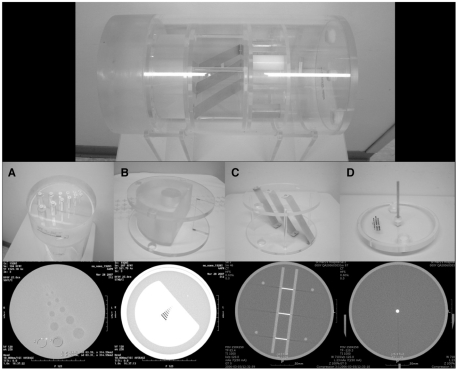
Fig. 1 AAPM CT performance phantom model 76-410. This photograph includes each phantom for evaluation of parameters and scanned images.A. Phantom for evaluation of low contrast resolutionB. Phantom for evaluation of spatial resoluationC. Phantom for evaluation of slice thicknessD. Phantom for evaluation of CT number of water, noise, and image uniformity
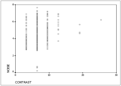
Fig. 2 Correlation between noise level and low contrast resolution. Correlation between noise and low contrast resolution showed statistically significant positive correlation (r = 0.183, p < 0.001, r = correlation coefficient).
Cited by 2 articles
-
Quantitative Analysis of the Effect of Iterative Reconstruction Using a Phantom: Determining the Appropriate Blending Percentage
Hyun Gi Kim, Yong Eun Chung, Young Han Lee, Jin-Young Choi, Mi-Suk Park, Myeong-Jin Kim, Ki Whang Kim
Yonsei Med J. 2015;56(1):253-261. doi: 10.3349/ymj.2015.56.1.253.Acceptance Test and Clinical Commissioning of CT Simulator
Hyun Joon An, Jaeman Son, Hyeongmin Jin, Jiwon Sung, Minsoo Chun
Prog Med Phys. 2019;30(4):160-166. doi: 10.14316/pmp.2019.30.4.160.
Reference
-
1. Reddinger Wil. CT image quality. 1998. OutSource, Inc.;http://www.e-radiography.net/mrict/CT_IQ.pdf.2. Im TH, Na DG. Annual report No. 1 2004~2006. 2007. Seoul: Korean Institute for Accreditation of Medical Image (KIAMI);p. 1–28. p. 75–105.3. Department of education, KIAMI. The workshop for the examiners on quality assurance program (2005). 2005. Seoul: KIAMI;p. 13–31.4. Department of education of KIAMI. The workshop for the examiners of quality assurance program (2006). 2006. Seoul: KIAMI;p. 141–177.5. McCollough CH, Bruesewitz MR, McNitt-Gray MF, Bush K, Ruckdeschel T, Payne JT, et al. The phantom portion of the American College of Radiology (ACR) computed tomography (CT) accreditation program: practical tips, artifact examples, and pitfalls to avoid. Med Phys. 2004; 31:2423–2442. PMID: 15487722.
Article6. Nuclear Associates 76-410-4130 and 76-411, AAPM CT performance phantom, users manual, Fluke Corporation. 2005. 3.7. Park HJ, Jung SE, Lee YJ, Cho WI, Do KH, Kim SH, et al. Review of failed CT phantom image evaluations in 2005 and 2006 by the CT accreditation program of the Korean Institute for Accreditation of Medical Image. Korean J Radiol. 2008; 9:354–363. PMID: 18682674.
Article8. CT accreditation program requirement. American Colleges of Radiology (ACR). http://www.acr.org/accreditation/computed/ct_reqs.aspx.9. National Council on Radiation protection and measurements. NCRP report No. 99. Quality assurance for diagnostic imaging equipment. 1988. Bethesda, Md: National Council on Radiation Protection and Measurements.10. McCollough CH, Zink FE. Goldman LW, editor. Performance evaluation of CT systems. Categorical course in diagnostic radiology physics: CT and US cross-sectional imaging. 2000. Oak Brook, IL:: RSNA;p. 189–207.11. Judy PF, Balter S, Bassano D, McCullough EC, Payne JT, Rothenberg L. AAPM report No. 1 phantoms of performance evaluation and quality assurance of CT scanner. 1977. Chicago, Illinois: AAPM.12. Euclid S. Computed tomography: physical principles, clinical applications and quality assurance. 1994. Philadelphia: Saunders;p. 174–199.
- Full Text Links
-
- Actions
-
Cited
- CITED
-
- Close
- Share
-
- Similar articles
-
- Objective and Quantitative Evaluation of Image Quality Using Fuzzy Integral: Phantom Study
- Review of Failed CT Phantom Image Evaluations in 2005 and 2006 by the CT Accreditation Program of the Korean Institute for Accreditation of Medical Image
- Enhancement of the Deformable Image Registration Accuracy Using Image Modification of MV CBCT
- Change of Radiologic Index of Foot according to Radiation Projection Angle: A Study Using Phantom Foot
- Optimization of exposure parameters and relationship between subjective and technical image quality in cone-beam computed tomography
