Ultrasonographic Characteristics of Subacute Granulomatous Thyroiditis
- Affiliations
-
- 1Department of Diagnostic Radiology, Gachon University Gil Medical Center, Incheon, Korea. ekkim@yumc.yonsei.ac.kra
- 2Department of Diagnostic Radiology, Yonsei University College of Medicine, Seoul, Korea.
- 3Department of Diagnostic Radiology, Sungkyunkwan University School of Medicine, Kangbuk Samsung Hospital, Seoul, Korea.
- 4Department of Pathology, Yonsei University College of Medicine, Seoul, Korea.
- 5Department of General Surgery, Yonsei University College of Medicine, Seoul, Korea.
- KMID: 1092545
- DOI: http://doi.org/10.3348/kjr.2006.7.4.229
Abstract
OBJECTIVE
We wanted to describe the characteristic ultrasonography (US) features and clinical findings for making the diagnosis of subacute granulomatous thyroiditis. MATERIALS AND METHODS: A total of 31 lesions from 27 patients were confirmed as subacute granulomatous thyroiditis by US-guided fine needle aspiration biopsy. We analyzed the ultrasonographic findings such as the lesion's size, margin and shape, the discrepancy between length and breadth and the vascularity. The clinical findings such as acute neck pain or fever were reviewed. The follow-up clinical and ultrasonographic data were reviewed for 15 patients. RESULTS: The thyroid gland was found to be enlarged in five patients, it was normal size in 20 patients and it was smaller in two patients. All the lesions had focally ill-defined hypoechogenicity. Hypervascularity was not noted in any of the lesions. Painful neck swelling was present in 18 patients. An accompanying fever was documented in nine of the 18 patients. Twelve patients showed disappearance (n = 3) or a decreased size (n = 9) of their lesions on follow-up US. CONCLUSION: The presence of ill-defined hypoechoic thyroid lesions without a discrete round or oval shape is characteristic for subacute granulomatous thyroiditis, and particularly when this is associated with painful neck swelling and/or fever.
Keyword
MeSH Terms
Figure
-
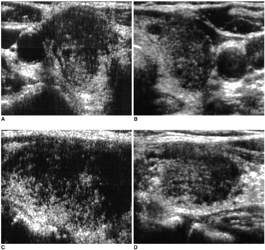
Fig. 1 A 62-year-old woman with diffuse neck swelling and malaise. The laboratory tests suggested normal thyroid function. The transverse right (A) and left (B), and longitudinal right (C) and left (D) thyroid sonograms show ill-defined hypoechoic lesions involving nearly the entire area of both thyroid glands. Both thyroids are diffusely enlarged, but no cervical lymphadenopathy was detected. Subacute granulomatous thyroiditis was confirmed by fine needle aspiration biopsy. The patient's condition improved dramatically following steroid treatment.
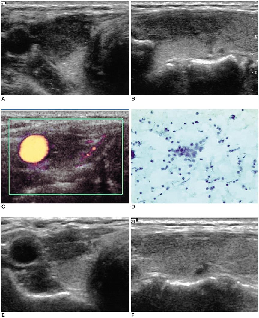
Fig. 2 A 45-year-old woman with severe neck pain and low-grade fever. The transverse (A) and longitudinal (B) sonograms of the right thyroid reveal an ill-defined elongated hypoechoic lesion, which is a typical finding of subacute thyroiditis. Color Doppler ultrasonography (C) shows no vascular flow in the hypoechoic lesion. Cytology suggests subacute granulomatous thyroiditis (D). On the day after steroid therapy, the patient felt free of neck pain. On sonogram after two months (E, F), the size of the hypoechoic area was markedly decreased.
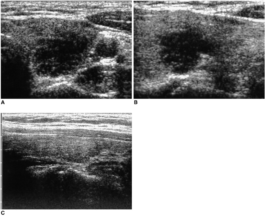
Fig. 3 A 50-year-old woman with neck swelling. Transverse (A) and longitudinal (B) sonograms of the left thyroid show an ill-defined, markedly hypoechoic lesion mimicking a malignant nodule. Subacute granulomatous thyroiditis was confirmed by performing fine needle aspiration biopsy. On the follow-up longitudinal sonogram after one month of medication (C), the lesion is not clearly visualized.
Cited by 2 articles
-
Painful Hashimoto Thyroiditis in a 7-Year-Old Girl: Differential Diagnosis and Medical Treatment
Gihong Park, Kyungchul Song, Hyun Joo Shin, Hyun Wook Chae
Int J Thyroidol. 2021;14(1):50-54. doi: 10.11106/ijt.2021.14.1.50.Thyroid Ultrasonography: Pitfalls and Techniques
Seon Hyeong Choi, Eun-Kyung Kim, Soo Jin Kim, Jin Young Kwak
Korean J Radiol. 2014;15(2):267-276. doi: 10.3348/kjr.2014.15.2.267.
Reference
-
1. Zacharia TT, Perumpallichira JJ, Sindhwani V, Chavhan G. Gray-scale and color Doppler sonographic findings in a case of subacute granulomatous thyroiditis mimicking thyroid carcinoma. J Clin Ultrasound. 2002. 30:442–444.2. Slatosky J, Shipton B, Wahba H. Thyroiditis: differential diagnosis and management. Am Fam Physician. 2000. 61:1047–1052. 10543. Benker G, Olbricht T, Windeck R, Wagner R, Albers H, Lederbogen S, et al. The sonographical and functional sequelae of de Quervain's subacute thyroiditis: long-term follow-up. Acta Endocrinol (Copenh). 1988. 117:435–441.4. Jhaveri K, Shroff MM, Fatterpekar GM, Som PM. CT and MR imaging findings associated with subacute thyroiditis. Am J Neuroradiol. 2003. 24:143–146.5. Qari FA. Tender neck in a diabetic patient. Saudi Med J. 2003. 24:675–676.6. Fatourechi V, Aniszewski JP, Fatourechi GZ, Atkinson EJ, Jacobsen SJ. Clinical features and outcome of subacute thyroiditis in an incidence cohort: Olmsted County, Minnesota, study. J Clin Endocrinol Metab. 2003. 88:2100–2105.7. Langer JE, Khan A, Nisenbaum HL, Baloch ZW, Horii SC, Coleman BG, et al. Sonographic appearance of focal thyroiditis. AJR Am J Roentgenol. 2001. 176:751–754.8. Vulpoi C, Zbranca E, Preda C, Ungureanu MC. [Contribution of ultrasonography in the evaluation of subacute thyroiditis]. Rev Med Chir Soc Med Nat Iasi. 2001. 105:749–755.9. Tokuda Y, Kasagi K, Iida Y, Yamamoto K, Hatabu H, Hidaka A, et al. Sonography of subacute thyroiditis: changes in the findings during the course of the disease. J Clin Ultrasound. 1990. 18:21–26.10. Brkljacic B, Cuk V, Tomic-Brzac H, Bence-Zigman Z, Delic-Brkljacic D, Drinkovic I. Ultrasonic evaluation of benign and malignant nodules in echographically multinodular thyroids. J Clin Ultrasound. 1994. 22:71–76.11. Aydin O, Apaydin FD, Bozdogan R, Pata C, Yalcinoglu O, Kanik A. Cytological correlation in patients who have a prediagnosis of thyroiditis ultrasonographically. Endocr Res. 2003. 29:97–106.12. Summaria V, Mirk P, Costantini AM, Maresca G, Ardito G, Bellantone R, et al. Role of Doppler color ultrasonography in the diagnosis of thyroid carcinoma. Ann Ital Chir. 2001. 72:277–282.
- Full Text Links
-
- Actions
-
Cited
- CITED
-
- Close
- Share
-
- Similar articles
-
- A Case of Graves' Disease Following Subacute Thyroiditis
- A Case of Painful Hashimoto's Thyroiditis Successfully Treated with Total Thyroidectomy
- Histopathologic study of the so called 'palpation thyroiditis'
- A Case of Riedel's Thyroiditis in a Patient with a History of Subacute Thyroiditis
- A case of an autonomously functioning thyroid nodule combined with subacute thyroiditis
