Hematology, cytochemistry and ultrastructure of blood cells in fishing cat (Felis viverrina)
- Affiliations
-
- 1Department of Pathology, Faculty of Veterinary Medicine, Kasetsart University, Bangkok-10900, Thailand. fvetksp@ku.ac.th
- 2Department of Pathology, Faculty of Medicine, Ramathibodi Hospital, Mahidol University, Bangkok-10400, Thailand.
- 3Central Instrumentation Unit, Faculty of Science, Mahasarakarm University, Mahasarakarm-44150, Thailand.
- KMID: 1089668
- DOI: http://doi.org/10.4142/jvs.2007.8.2.163
Abstract
- Hematological, cytochemical and ultrastructural features of blood cells in fishing cat (Felis viverrina) were evaluated using complete blood cell counts with routine and cytochemical blood stains, and scanning and transmission electron microscopy. No statistically significant difference was found in different genders of this animal. Unique features of blood cells in this animal were identified in hematological, cytochemical and ultrastructural studies. This study contributes to broaden hematological resources in wildlife animals and provides a guideline for identification of blood cells in the fishing cat.
Keyword
MeSH Terms
Figure
-
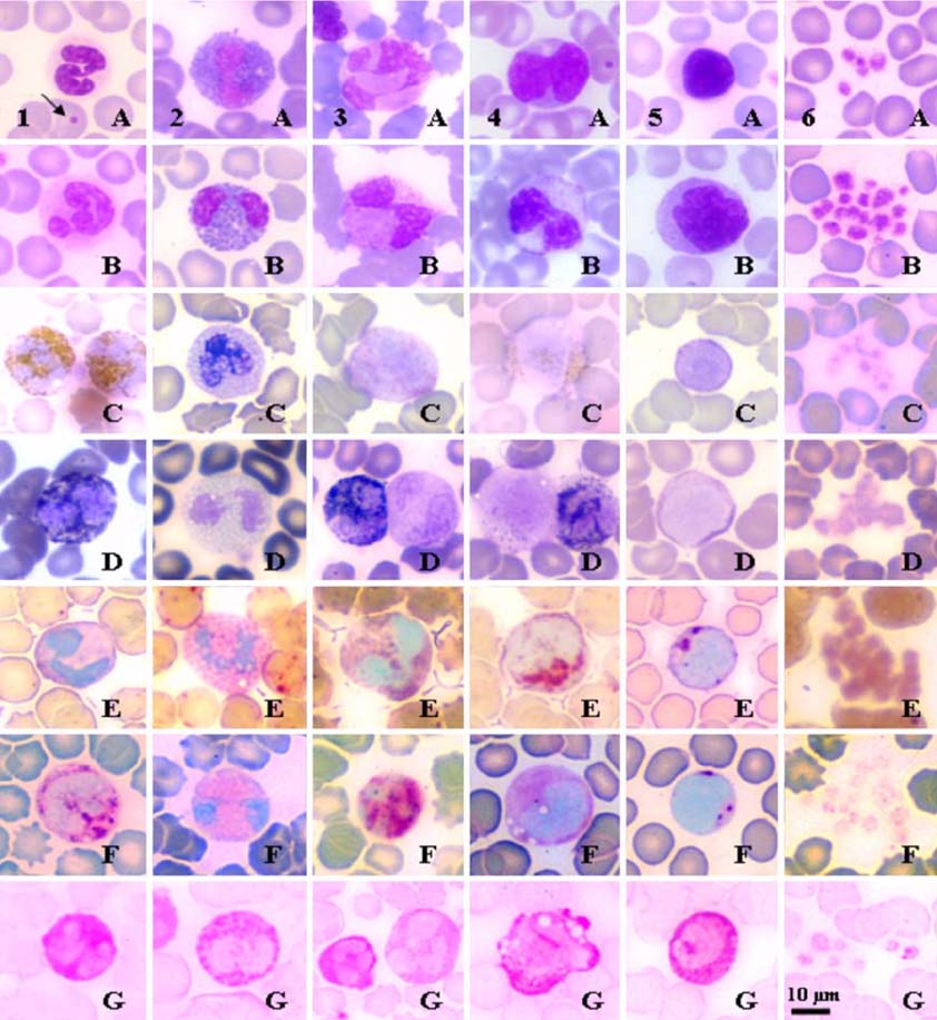
Fig. 1 to 6 Romanowsky's and selected cytochemical staining reactions for normal blood cells in fishing cat. Fig. 1. Neutrophils: (A & B) a constricted nucleus with indiscernible cytoplasmic granules, one Howell-Jolly bodies nearby (arrow), WG and W, respectively. (C & D) strongly positive neutrophils, PER and SBB, respectively. (E) weakly positive for ANAE. (F) positively stained for β-glu. (G) strongly positive for PAS. Fig. 2. Eosinophils: (A & B) an eosinophil with rod-shape granules, WG and W, respectively. (C & D) refractive granules in non stained cytoplasm, PER and SBB, respectively. (E) refractive granules in red-brown cytoplasm, ANAE. (F) positive in the periphery of the granules, β-glu. (G) non-stained granules in strong magenta cytoplasm, PAS. Fig. 3. Basophils: (A) a basophil with large lobed nucleus and lavender-stained cytoplasm with WG. (B) a few dark purple granules in cytoplasm with W. (C) negative for PER. (D) a negatively stained basophil (right) compared to a positively stained neutrophil (left), SBB. (E & F) positive for ANAE and β-glu, respectively. (G) positive with fine granular pattern of basophil (right) compared to positive with diffuse pattern of neutrophil (left), PAS. Fig. 4. Monocytes: (A & B) a monocyte with deep indented nucleus, WG and W, respectively. (C) slightly positive for PER. (D) slightly positive with a few black granules of monocyte (left) compared to strongly positive neutrophil (right), SBB. (E, F & G) positive for ANAE, β-glu and PAS, respectively. Fig. 5. Lymphocytes: (A & B) a lymphocyte with condense nuclear chromatin and thin band cytoplasm, WG and W, respectively. (C & D) negative for PER and SBB, respectively. (E, F & G) positive for ANAE, β-glu and PAS, respectively. Fig. 6. Platelets: (A & B) group of small round, anucleated cells with reddish purple granules, WG and W, respectively. (C & D) negative for PER and SBB, respectively. (E) strongly positive for ANAE. (F & G) weakly positive for β-glu and PAS, respectively.
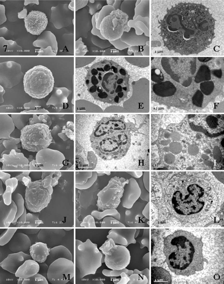
Fig. 7 Cellular surfaces and ultrastructures of various white blood cells in fishing cat. (A) a neutrophil showing numerous microvilli on its surface in contact with one platelet, SEM. (B) a neutrophil with ruffled membrane protuberance, SEM. (C) a neutrophil presenting short microvilli, lobulated nucleus, cytoplasmic granules and vesicular bodies, TEM. (D) an eosinophil showing custard-apple liked appearance of granule contour with microvilli, SEM. (E) an eosinophil containing characteristic large granules, TEM. (F) higher magnification of (E), presenting round to angulated electron dense granules ranging 0.5-1 µm in diameter. (G) a basophil revealed irregular rod shaped granule contour, SEM. (H) a basophil showing large lobed nucleus with heterogenous granules, membrane folds, microvilli and irregular branch projection, TEM. (I) higher magnification of (H) showing basophil granules. (J) a monocyte showing wavy appearance with pseudopodic projections, SEM. (K) a monocyte showing lamellipodium extended from the cell center, SEM. (L) a monocyte showing deep indented nucleus and less electron dense cytoplasm, TEM. (M) a lymphocyte presenting bulgy contour with several blebs, SEM. (N) a lymphocyte showing lamellipodium extended from cell margin, SEM. (O) a lymphocyte revealed nuclear indentation with cell membrane folds and a few microvillous projections, TEM.
Reference
-
1. Clinkenbeard KD, Meinkoth JH. Feldman BF, Zinkl JG, Jain NC, editors. Normal hematology of the cat. Schalm's Veterinary Hematology. 2000. 5th ed. Philadelphia: Lippincott Williams & Wilkins;1064–1068.2. Hayhoe FGJ, Quaglino D. Haematological Cytochemistry. 1980. Edinburgh: Churchill Livingstone;68–75.3. Jain NC. Essentials of Veterinary Hematology. 1993. Philadelphia: Lea & Febiger;417.4. Jain NC. Schalm's Veterinary Hematology. 1986. 4th ed. Philadelphia: Lea & Febiger;1221.5. Kaplow LS. Simplified myeloperoxidase stain using benzidine dihydrochloride. Blood. 1965. 26:215–219.6. Lekagul B, McNeely JA, editors. Family felidae. Mammals of Thailand. 1988. 2nd ed. Bangkok: Darnsutha Press;603–630.7. Raskin RE, Valenciano A. Feldman BF, Zinkl JG, Jain NC, editors. Cytochemistry of normal leukocytes. Schalm's Veterinary Hematology. 2000. 5th ed. Philadelphia: Lippincott Williams & Wilkins;337–346.8. Reagan WJ, Sanders TG, DeNicola DB. Veterinary Hematology: Atlas of Common Domestic Species. 1998. Ames: Manson Publishing;75.9. Salakij C, Salakij J, Apibal S, Narkkong NA, Chanhome L, Rochanapat N. Hematology, morphology, cytochemical staining, and ultrastructural characteristics of blood cells in king cobras (Ophiophagus hannah). Vet Clin Pathol. 2002. 31:116–126.
Article10. Salakij C, Salakij J, Narkkong NA, Trongwonsa L, Pattanarangsan R. Hematology, cytochemistry and ultrastructure of blood cells from Asiatic black bear (Ursus thibetanus). Kasetsart J (Nat Sci). 2005. 39:247–261.11. Salakij J, Salakij C, Narkkong NA, Apibal S, Suthunmapinuntra P, Rattanakunuprakarn J, Nunklang G, Yindee M. Hematology, cytochemistry and ultrastructure of blood cells from Asian elephant (Elephas maximus). Kasetsart J (Nat Sci). 2005. 39:482–493.12. Sheehan HL, Storey GW. An improved method of staining leukocyte granules with Sudan black B. J Pathol Bacteriol. 1947. 59:336–337.13. Steffens WL. Feldman BF, Zinkl JG, Jain NC, editors. Ultrastructure features of leukocytes. Schalm's Veterinary Hematology. 2000. 5th ed. Philadelphia: Lippincott Williams & Wilkins;326–336.14. Tablin F. Feldman BF, Zinkl JG, Jain NC, editors. Platelet Structure and Function. Schalm's Veterinary Hematology. 2000. 5th ed. Philadelphia: Lippincott Williams & Wilkins;448–452.15. Tsujimoto H, Hasegawa A, Tomoda I. A cytochemical study on feline blood cells. Nippon Juigaku Zasshi. 1983. 45:373–382.
Article16. Yam LT, Li CY, Crosby WH. Cytochemical identification of monocytes and granulocytes. Am J Clin Pathol. 1971. 55:283–290.
Article
- Full Text Links
-
- Actions
-
Cited
- CITED
-
- Close
- Share
-
- Similar articles
-
- Acute monoblastic leukemia in a FeLV-positive cat
- Surface ultrastructure of the adult stage of Acanthotrema felis (Trematoda: Heterophyidae
- Evaluation of assays to detect Helicobacter felis infection in cats
- Detection of Helicobacter felis in a cat with gastric disease in laboratory animal facility
- Investigation of chlamydophilosis from naturally infected cats
