Acute monoblastic leukemia in a FeLV-positive cat
- Affiliations
-
- 1Department of Pathology, Faculty of Veterinary Medicine, Kasetsart University, Bangkok-10900, Thailand. fvetksp@ku.ac.th
- 2Central Instrumentation Unit, Faculty of Science, Mahasarakarm University, Mahasarakarm-44150, Thailand.
- 3Department of Pathology, Faculty of Medicine, Ramathibodi Hospital, Mahidol University, Bangkok-10400, Thailand.
- KMID: 1102961
- DOI: http://doi.org/10.4142/jvs.2008.9.1.109
Abstract
- A 1.6-year-old male domestic short hair cat was brought to the Veterinary Medical Teaching Hospital, Kasetsart University, with signs of severe anemia, depression, and general lymph node enlargement. Complete blood count revealed leukocytosis and massive undifferentiated blasts. Testing for antibodies specific to feline leukemia virus (FeLV) was positive, and FeLV nucleic acid was confirmed by nested polymerase chain reaction. Base on cytochemistry and ultrastructure, the cat was diagnosed with acute monoblastic leukemia.
Keyword
MeSH Terms
Figure
-
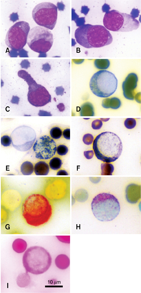
Fig. 1 Morphologies and cytochemical stainings of undifferentiated blasts: (A and B) blasts were round to ovoid in shape with finely stippled nuclear chromatin and distinct nucleoli (Wright-Giemsa stain); (C) a blast with a prominent cytoplasmic tail (Wright-Giemsa stain); (D) a blast that stained positive for peroxidase; (E) a blast that was negatively stained for Sudan black B (left) compared to a positively-stained granulocyte (right); (F) a blast with positive Sudan black B staining; (G) a blast that was strongly positive for α-naphthyl acetate esterase; (H) a blast that was moderately positive for β-glucuronidase; (I) a blast that was positive for PAS.
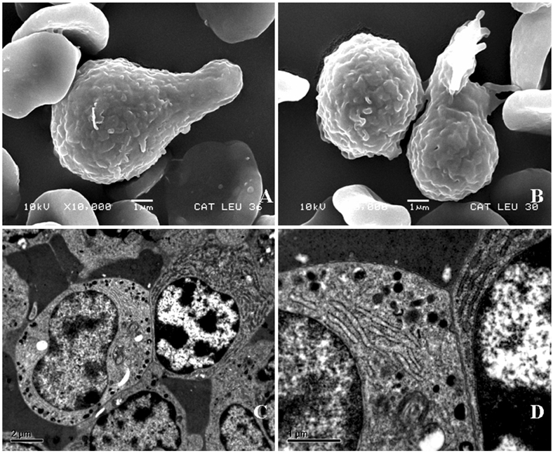
Fig. 2 Cellular surfaces and ultrastructures of blasts: (A and B) Ruffled membrane with deep fissures and pseudopodic projection (SEM); (C) Indented nucleus with marginated chromatin and light cytoplasm with some electron dense granules and organelles (left) adhered to a plasma cell (right) (TEM); (D) High magnification of (C) showing the organelles and granules.
Reference
-
1. Grindem CB. Ultrastructural morphology of leukemic cells from 14 dogs. Vet Pathol. 1985. 22:456–462.
Article2. Grindem CB, Stevens JB, Perman V. Cytochemical reactions in cells from leukemic dogs. Vet Pathol. 1986. 23:103–109.
Article3. Hayhoe FGJ, Quaglino D. Hematological Cytochemistry. 1980. Edinburgh: Churchill Livingstone;68–75.4. Jain NC. Schalm's Veterinary Hematology. 1986. 4th ed. Philadelphia: Lea & Febiger;1221.5. Kass L. Leukemia, Cytology and Cytochemistry. 1982. Philadelphia: Lippincott;167–188.6. Khan KNM, Kociba GJ, Wellman ML. Macrophage tropism of feline leukemia virus (FeLV) of subgroup-C and increased production of tumor necrosis factor-α by FeLV-infected macrophages. Blood. 1993. 81:2585–2590.
Article7. Kohlmann A, Schoch C, Dugas M, Schnittger S, Hiddemann W, Kern W, Haferlach T. New insights into MLL gene rearranged acute leukemias using gene expression profiling: shared pathways, lineage commitment, and partner genes. Leukemia. 2005. 19:953–964.
Article8. Kuwada N, Kimura F, Matsumura T, Yamashita T, Nakamura Y, Wakimoto N, Ikeda T, Sato K, Motoyoshi K. t(11;14)(q23;q24) generates an MLL-human gephyrin fusion gene along with a de facto truncated MLL in acute monoblastic leukemia. Cancer Res. 2001. 61:2665–2669.9. Miyazawa T, Jarrett O. Feline leukaemia virus proviral DNA detected by polymerase chain reaction in antigenaemic but non-viraemic ('discordant') cats. Arch Virol. 1997. 142:323–332.
Article10. Prihirunkit K, Kasorndokbua C, Apibal S. Diagnosis of acute lymphoblastic leukemia in a dog by cytochemistry. J Thai Vet Pract. 2005. 17:53–61.11. Salakij C, Salakij J, Apibal S, Narkkong NA, Chanhome L, Rochanapat N. Hematology, morphology, cytochemical staining, and ultrastructural characteristics of blood cells in king cobras (Ophiophagus hannah). Vet Clin Pathol. 2002. 31:116–126.
Article12. Thrall MA. Lymphoproliferative disorders: lymphocytic leukemia and plasma cell myeloma. Vet Clin North Am Small Anim Pract. 1981. 11:321–347.
- Full Text Links
-
- Actions
-
Cited
- CITED
-
- Close
- Share
-
- Similar articles
-
- A Case of Acute Monoblastic Leukemia with Hexasomy 8
- A Case of Bone Marrow Necrosis Preceeding Acute Monoblastic Leukemia
- A Case of Acute Monoblastic Leukemia developed during Pregnancy
- Acute Monoblastic Leukemia with t(11;17)(q23;q21): Fusion of the KMT2A(MLL) and MLLT6(AF17) Genes
- A Rare Case of Cutaneous T-Cell Lymphoma Accompanied by Acute Monoblastic Leukemia and Diffuse Large B-Cell Lymphoma
