Reconstruction Algorithms Influence the Follow-Up Variability in the Longitudinal CT Emphysema Index Measurements
- Affiliations
-
- 1Universidade Federal do Rio de Janeiro, Radiology Department, Av. Pedro Calmon, ndegrees 550 - Cidade Universitaria, 21941-901, Rio de Janeiro, RJ, Brazil.
- 2Moinhos de Vento Hospital, Chest Radiology Department, Rua Tiradentes 333, Bairro Moinhos de Vento, 90050-123, Porto Alegre, RS, Brazil.
- 3Post-Graduation Program in Medicine, Chest Medicine, the Federal University of Rio Grande do Sul, Rua Ramiro Barcelos, 2400, 2degrees andar, 90035-003, Porto Alegre, RS, Brazil.
- 4Liverpool Heart and Chest Hospital NHS Foundation Trust, Thomas Drive, Liverpool L14 3PE, England.
- KMID: 1088561
- DOI: http://doi.org/10.3348/kjr.2011.12.2.169
Abstract
OBJECTIVE
We wanted to compare the variability in the longitudinal emphysema index (EI) measurements that were computed with standard and high resolution (HR) reconstruction algorithms (RAs).
MATERIALS AND METHODS
We performed a retrospective review of 475 patients who underwent CT for surveillance of lung nodules. From this cohort, 50 patients (28 male) were included in the study. For these patients, the baseline and follow-up scans were acquired on the same multidetector CT scanner and using the same acquisition protocol. The CT scans were reconstructed with HR and standard RAs. We determined the difference in the EI between CT1 and CT2 for the HR and standard RAs, and we compared the variance of these differences.
RESULTS
The mean of the variation of the total lung volume was 0.14 L (standard deviation [SD] = 0.13 L) for the standard RA and 0.16 L (SD = 0.15 L) for the HR RA. These differences were not significant. For the standard RA, the mean variation was 0.13% (SD = 0.44%) for EI -970 and 0.4% (SD = 0.88%) for EI -950; for the HR RA, the mean variation was 1.9% (SD = 2.2%) for EI -970 and 3.6% (SD = 3.7%) for EI -950. These differences were significant.
CONCLUSION
Using an HR RA appears to increase the variability of the CT measurements of the EI.
MeSH Terms
Figure
-
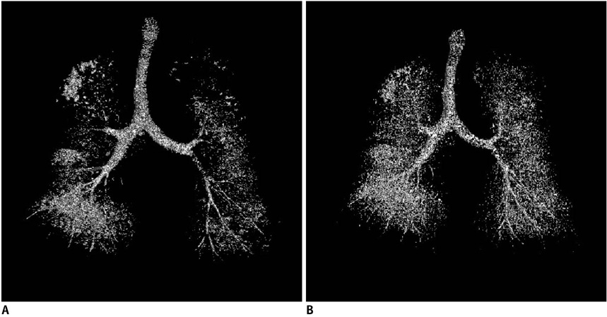
Fig. 1 3D CT volume with emphysema volumes in white. A. Reconstruction image with standard algorithm. B. Reconstruction image with high resolution algorithm. Note difference in emphysema volumes.
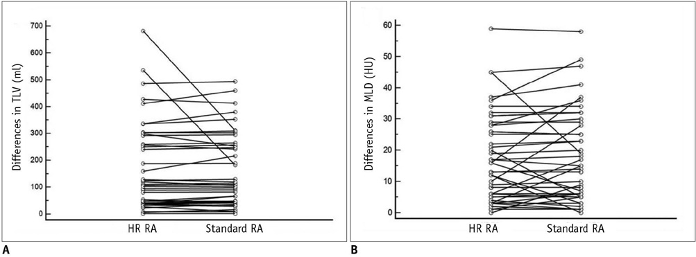
Fig. 2 Comparison of differencs in CT measurements of total lung volume at CT1 and CT2 between reconstruction algorithm. A. Comparison of differences in CT measurements of total lung volume (TLV) at CT1 and CT2 between high resolution (HR) reconstruction algorithm (RA) and standard reconstruction algorithm. B. Comparison of differences in CT measurements of mean lung density (MLD) at CT1 and CT2 between high resolution reconstruction algorithm and standard reconstruction algorithm.
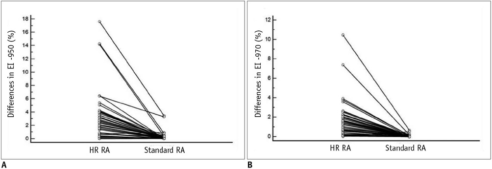
Fig. 3 Comparison of differences in CT measurements of EI -950 at CT1 and CT2 between reconstruction algorithm. A. Comparison of differences in CT measurements of EI -950 at CT1 and CT2 between high resolution (HR) reconstruction algorithm (RA) and standard reconstruction algorithm. B. Comparison of differences in CT measurements of EI -970 at CT1 and CT2 between high resolution reconstruction algorithm and standard reconstruction algorithm.
Reference
-
1. The definition of emphysema. Report of a National Heart, Lung, and Blood Institute, Division of Lung Diseases workshop. Am Rev Respir Dis. 1985. 132:182–185.2. Park SO, Seo JB, Kim N, Park SH, Lee YK, Park BW, et al. Feasibility of automated quantification of regional disease patterns depicted on high-resolution computed tomography in patients with various diffuse lung diseases. Korean J Radiol. 2009. 10:455–463.3. Newell JD Jr, Hogg JC, Snider GL. Report of a workshop: quantitative computed tomography scanning in longitudinal studies of emphysema. Eur Respir J. 2004. 23:769–775.4. Irion KL, Hochhegger B, Marchiori E, Porto Nda S, Baldisserotto Sde V, Santana PR. Chest X-ray and computed tomography in the evaluation of pulmonary emphysema. J Bras Pneumol. 2007. 33:720–732.5. Kemerink GJ, Kruize HH, Lamers RJ, van Engelshoven JM. Density resolution in quantitative computed tomography of foam and lung. Med Phys. 1996. 23:1697–1708.6. Boedeker KL, McNitt-Gray MF, Rogers SR, Truong DA, Brown MS, Gjertson DW, et al. Emphysema: effect of reconstruction algorithm on CT imaging measures. Radiology. 2004. 232:295–301.7. Ley-Zaporozhan J, Ley S, Weinheimer O, Iliyushenko S, Erdugan S, Eberhardt R, et al. Quantitative analysis of emphysema in 3D using MDCT: influence of different reconstruction algorithms. Eur J Radiol. 2008. 65:228–234.8. Parr DG, Stoel BC, Stolk J, Stockley RA. Validation of computed tomographic lung densitometry for monitoring emphysema in alpha1-antitrypsin deficiency. Thorax. 2006. 61:485–490.9. Dowson LJ, Newall C, Guest PJ, Hill SL, Stockley RA. Exercise capacity predicts health status in alpha(1)-antitrypsin deficiency. Am J Respir Crit Care Med. 2001. 163:936–941.10. Dirksen A, Piitulainen E, Parr DG, Deng C, Wencker M, Shaker SB, et al. Exploring the role of CT densitometry: a randomised study of augmentation therapy in alpha1-antitrypsin deficiency. Eur Respir J. 2009. 33:1345–1353.11. Desai SR, Hansell DM, Walker A, MacDonald SL, Chabat F, Wells AU. Quantification of emphysema: a composite physiologic index derived from CT estimation of disease extent. Eur Radiol. 2007. 17:911–918.12. Kim WJ, Silverman EK, Hoffman E, Criner GJ, Mosenifar Z, Sciurba FC, et al. CT metrics of airway disease and emphysema in severe COPD. Chest. 2009. 136:396–404.13. Ogawa E, Nakano Y, Ohara T, Muro S, Hirai T, Sato S, et al. Body mass index in male patients with COPD: correlation with low attenuation areas on CT. Thorax. 2009. 64:20–25.14. Goo JM, Kim KG, Gierada DS, Castro M, Bae KT. Volumetric measurements of lung nodules with multi-detector row CT: effect of changes in lung volume. Korean J Radiol. 2006. 7:243–248.15. Gevenois PA, De Vuyst P, de Maertelaer V, Zanen J, Jacobovitz D, Cosio MG, et al. Comparison of computed density and microscopic morphometry in pulmonary emphysema. Am J Respir Crit Care Med. 1996. 154:187–192.16. Madani A, Zanen J, de Maertelaer V, Gevenois PA. Pulmonary emphysema: objective quantification at multi-detector row CT--comparison with macroscopic and microscopic morphometry. Radiology. 2006. 238:1036–1043.17. Gevenois PA, Yernault JC. Can computed tomography quantify pulmonary emphysema? Eur Respir J. 1995. 8:843–848.18. Litmanovich D, Boiselle PM, Bankier AA. CT of pulmonary emphysema--current status, challenges, and future directions. Eur Radiol. 2009. 19:537–551.19. Rosenblum LJ, Mauceri RA, Wellenstein DE, Bassano DA, Cohen WN, Heitzman ER. Computed tomography of the lung. Radiology. 1978. 129:521–524.20. Hayhurst MD, MacNee W, Flenley DC, Wright D, McLean A, Lamb D, et al. Diagnosis of pulmonary emphysema by computerised tomography. Lancet. 1984. 2:320–322.21. Muller NL, Staples CA, Miller RR, Abboud RT. "Density mask". An objective method to quantitate emphysema using computed tomography. Chest. 1988. 94:782–787.22. Shaker SB, Dirksen A, Laursen LC, Maltbaek N, Christensen L, Sander U, et al. Short-term reproducibility of computed tomography-based lung density measurements in alpha-1 antitrypsin deficiency and smokers with emphysema. Acta Radiol. 2004. 45:424–430.23. Omori H, Nakashima R, Otsuka N, Mishima Y, Tomiguchi S, Narimatsu A, et al. Emphysema detected by lung cancer screening with low-dose spiral CT: prevalence, and correlation with smoking habits and pulmonary function in Japanese male subjects. Respirology. 2006. 11:205–210.24. Schilham AM, van Ginneken B, Gietema H, Prokop M. Local noise weighted filtering for emphysema scoring of low-dose CT images. IEEE Trans Med Imaging. 2006. 25:451–463.25. Stoel BC, Putter H, Bakker ME, Dirksen A, Stockley RA, Piitulainen E, et al. Volume correction in computed tomography densitometry for follow-up studies on pulmonary emphysema. Proc Am Thorac Soc. 2008. 5:919–924.26. Gietema HA, Schilham AM, van Ginneken B, van Klaveren RJ, Lammers JW, Prokop M. Monitoring of smoking-induced emphysema with CT in a lung cancer screening setting: detection of real increase in extent of emphysema. Radiology. 2007. 244:890–897.
- Full Text Links
-
- Actions
-
Cited
- CITED
-
- Close
- Share
-
- Similar articles
-
- Impact of Model-Based Iterative Reconstruction on the Correlation between Computed Tomography Quantification of a Low Lung Attenuation Area and Airway Measurements and Pulmonary Function Test Results in Normal Subjects
- Clinical use of chest CT in chronic obstructive pulmonary diseases
- Comparison of Filtered Back Projection, Hybrid Iterative Reconstruction, Model-Based Iterative Reconstruction, and Virtual Monoenergetic Reconstruction Images at Both Low- and Standard-Dose Settings in Measurement of Emphysema Volume and Airway Wall Thickness: A CT Phantom Study
- The Influence of Reconstruction Algorithm and Heart Rate on Coronary Artery Image Quality and Stenosis Detection at 64-Detector Cardiac CT
- Pulmonary Emphysema: Visual Interpretation and Quantitative Analysis
