CT Arthrography and Virtual Arthroscopy in the Diagnosis of the Anterior Cruciate Ligament and Meniscal Abnormalities of the Knee Joint
- Affiliations
-
- 1Department of Radiology, Seoul National University College of Medicine the Institute of Radiation Medicine, SNUMRC. hongsh@radiol.snu.ac.kr
- 2Aeromedical Center, Republic of Korea Air Force.
- KMID: 1066245
- DOI: http://doi.org/10.3348/kjr.2004.5.1.47
Abstract
OBJECTIVE
To determine the diagnostic accuracy of CT arthrography and virtual arthroscopy in the diagnosis of anterior cruciate ligament and meniscus pathology. MATERIALS AND METHODS: Thirty-eight consecutive patients who underwent CT arthrography and arthroscopy of the knee were included in this study. The ages of the patients ranged from 19 to 52 years and all of the patients were male. Sagittal, coronal, transverse and oblique coronal multiplanar reconstruction images were reformatted from CT arthrography. Virtual arthroscopy was performed from 6 standard views using a volume rendering technique. Three radiologists analyzed the MPR images and two orthopedic surgeons analyzed the virtual arthroscopic images. RESULTS: The sensitivity and specificity of CT arthrography for the diagnosis of anterior cruciate ligament abnormalities were 87.5%-100% and 93.3-96.7%, respectively, and those for meniscus abnormalities were 91.7%-100% and 98.1%, respectively. The sensitivity and specificity of virtual arthroscopy for the diagnosis of anterior cruciate ligament abnormalities were 87.5% and 83.3-90%, respectively, and those for meniscus abnormalities were 83.3%-87.5% and 96.1-98.1%, respectively. CONCLUSION: CT arthrography and virtual arthroscopy showed good diagnostic accuracy for anterior cruciate ligament and meniscal abnormalities.
MeSH Terms
Figure
-
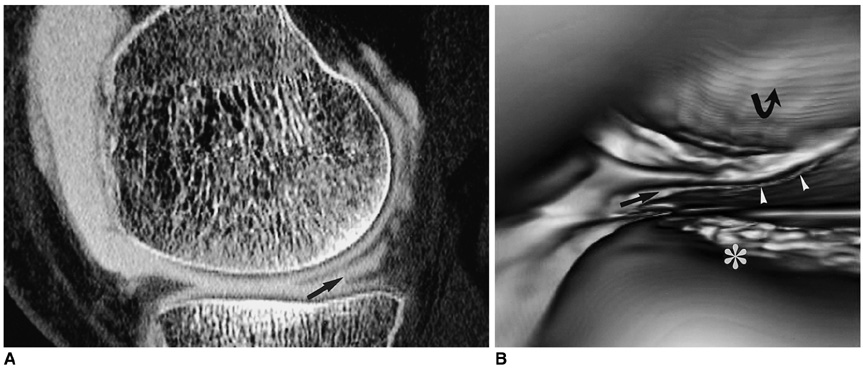
Fig. 1 Medial meniscus tear in a 28-year-old man with knee pain after trauma. A. Sagittal CT image clearly demonstrating an oblique tear of the medial meniscus (arrow). B. Virtual arthroscopy of the medial meniscus showing cleavage (arrow) and a rugged free margin of torn meniscus (arrowheads). The tibial plateau (asterisk) and femur condyle (curved arrow) are also shown.
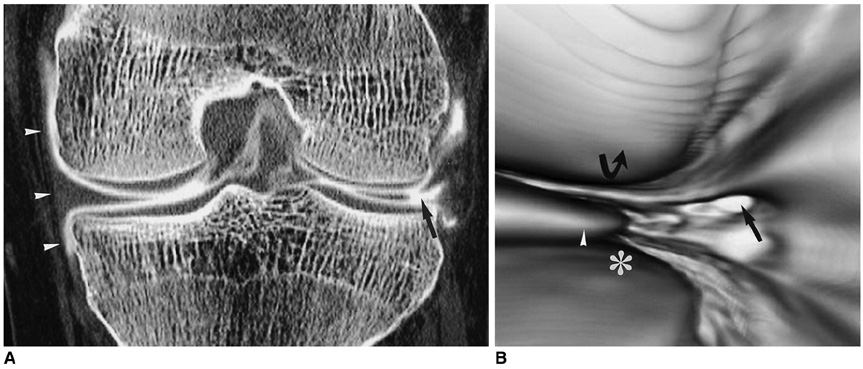
Fig. 2 CT arthrography and virtual arthroscopy of the lateral meniscus tear of a 29-year-old man with knee joint pain. A. Coronal CT arthrography showing a vertical and horizontal tear of the lateral meniscus (arrow). Note the intact medial collateral ligament (arrowheads). B. Virtual arthroscopy showing cleavage of the meniscus (arrow) and a displaced meniscus central segment (arrowhead). The tibia plateau (asterisk) and femur condyle (curved arrow) are clearly demonstrated.
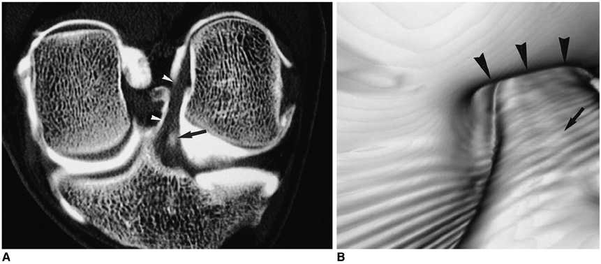
Fig. 3 CT arthrography of the normal anterior cruciate ligament of a 27-year-old male. A. Oblique coronal CT arthrography showing a well demarcated linear anterior cruciate ligament (arrowheads). The small amount of contrast media on the tibial side was considered a normal finding (arrow). B. Virtual arthroscopic view of the normal anterior cruciate ligament (arrow). The smooth margin and continuity to the intercondylar notch are clearly demonstrated (arrowheads).
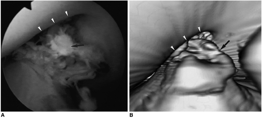
Fig. 4 A tear in the anterior cruciate ligament of a 20-year-old man with knee joint pain after trauma A. Arthroscopy showing a rugged torn fiber of the anterior cruciate ligament. One arthroscopist judged that there was a 50% partial tear of the anterior cruciate ligament. The ridge of the intercondylar notch (arrowheads) and the torn anterior cruciate ligament (arrow) are shown. B. Virtual arthroscopy showing an anterior cruciate ligament with abnormal shape and orientation. The ridge of the intercondylar notch is shown (arrowheads). The anterior cruciate ligament has lost its normal continuity and has an irregular torn margin (arrow).
Reference
-
1. Tavernier T, Dejour D. Knee imaging: what is the best modality. J Radiol. 2001. 82:387–408.2. Crues JV III, Mink J, Levy TL, Lotysch M, Stoller DW. Meniscal tears of the knee: accuracy of MR imaging. Radiology. 1987. 164:445–448.3. Mink JH, Levy T, Crues JV III. Tears of the anterior cruciate ligament and menisci of the knee: MR imaging evaluation. Radiology. 1988. 167:769–774.4. Ghelman B. Meniscal tears of the knee: evaluation by high-resolution CT combined with arthrography. Radiology. 1985. 157:23–27.5. Malghem J, Vande Berg BC, Lebon C, Lecouvet FE, Maldague BE. Ganglion cysts of the knee: articular communication revealed by delayed radiography and CT after arthrography. AJR Am J Roentgenol. 1998. 170:1579–1583.6. Berland LL, Smith JK. Multi detector-array CT: once again, technology creates new opportunities. Radiology. 1998. 209:327–329.7. Vande Berg BC, Lecouvet FE, Poilvache P, et al. Dual-detector spiral CT arthrography of the knee: accuracy for detection of meniscal abnormalities and unstable meniscal tear. Radiology. 2000. 216:851–857.8. Vande Berg BC, Lecouvet FE, Poilvache P, Maldague B, Malghem J. Spiral CT arthrography of the knee: technique and value in the assessment of internal derangement of the knee. Eur Radiol. 2002. 12:1800–1810.9. Irie K, Yamada T. Three-dimensional virtual computed tomography imaging for injured anterior cruciate ligament. Arch Orthop Trauma Surg. 2002. 122:93–95.10. Vande Berg BC, Lecouvet FE, Poilvache P, Dubuc JE, Maldague B, Malghem J. Anterior cruciate ligament tears and associated meniscal lesions: assessment at dual-detector spiral CT arthrography. Radiology. 2002. 223:403–409.11. Rubin DA. MR imaging of the knee menisci. Radiol Clin North Am. 1997. 35:21–44.12. Heron CW, Calvert PT. Three-dimensional gradient-echo MR imaging of the knee: comparison with arthroscopy in 100 patients. Radiology. 1992. 183:839–844.13. Pedowitz RA, Esch J, Snyder S. Evaluation of a virtual reality simulator for arthroscopy skills development. Arthroscopy. 2002. 18:29E.14. Mabrey JD, Gillogly SD, Kasser JR, Sweeney HJ, Zarins B, Mevis H. Virtual reality simulation of arthroscopy of the knee. Arthroscopy. 2002. 18:28E.
- Full Text Links
-
- Actions
-
Cited
- CITED
-
- Close
- Share
-
- Similar articles
-
- Computed tomography of the knee joint
- The Reconstruction of Anterior Cruciate Ligament Using Patellar Tendon under Arthroscopy
- Diagnostic Significance of Arthrography for the Internal Derangement of Knee Joint
- Technical evolution of arthoscopic knee surgery
- Internal derangement of the knee:Diagnostic accuracy of MR imaging
