Coronary Vasospastic Angina: Assessment by Multidetector CT Coronary Angiography
- Affiliations
-
- 1Division of Cardiovascular Imaging, Department of Radiology, Cardiovascular Center Seoul National University Bundang Hospital, Gyeonggi-do 436-707, Korea. drsic@radiol.snu.ac.kr
- 2Division of Cardiology, Department of Internal Medicine, Cardiovascular Center Seoul National University Bundang Hospital, Gyeonggi-do 436-707, Korea.
- KMID: 1058790
- DOI: http://doi.org/10.3348/kjr.2012.13.1.27
Abstract
OBJECTIVE
We aimed to describe the imaging findings of multidetector CT coronary angiography (MDCTA) in cases of vasospastic angina (VA) and to determine the accuracy of MDCTA in the identification of VA as compared with invasive coronary angiography with an ergonovine provocation test (CAG with an EG test).
MATERIALS AND METHODS
Fifty-three patients with clinically suspected VA were enrolled in this study. Two radiologists analyzed the stenosis degree, presence or absence of plaque, plaque composition, and a remodeling index of the related-segment in CAG with an EG test, which were used as a gold standard. We evaluated the diagnostic performances of MDCTA by comparing the MDCTA findings with those of CAG with an EG test.
RESULTS
Among the 25 patients with positive CAG with an EG test, all 12 patients with significant stenosis showed no definite plaque with the negative arterial remodeling. Of the six patients with insignificant stenosis, three (50%) had non-calcified plaque (NCP), two (33%) had mixed plaque, and one (17%) had calcified plaque. When the criteria for significant stenosis with negative remodeling but no definite evidence of plaque as a characteristic finding of MDCTA were used, results showed sensitivities, specificities, positive predictive values (PPV), and negative predictive values (NPV) of 48%, 100%, 100%, and 68%, respectively.
CONCLUSION
Significant stenosis with negative remodeling, but no definite evidence of plaque, is the characteristic finding on MDCTA of VA. Cardiac MDCTA shows good diagnostic performance with high specificity and PPV as compared with CAG with an EG test.
Keyword
MeSH Terms
-
Angina Pectoris/*radiography
Chi-Square Distribution
Comorbidity
Contrast Media/diagnostic use
Coronary Angiography/*methods
Electrocardiography
Ergonovine/diagnostic use
Female
Humans
Iopamidol/analogs & derivatives/diagnostic use
Male
Middle Aged
Oxytocics/diagnostic use
Predictive Value of Tests
Radiographic Image Interpretation, Computer-Assisted
Retrospective Studies
Sensitivity and Specificity
Tomography, X-Ray Computed/*methods
Figure
-
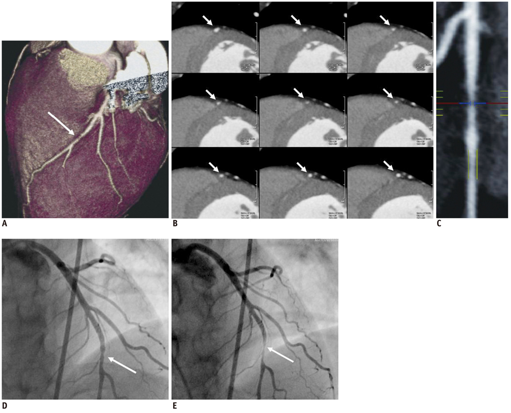
Fig. 1 41-year-old man with acute chest pain. Significant discrete stenosis is noted in middle segment of left anterior descending artery (arrows) on volume rendering image (A), short-axis multiplanar image (B), and curved multiplanar multidetector CT coronary angiography images (C). However, there is no discernable atheroma in related segment, suggesting coronary spasm (B). Mild luminal irregularity is shown at corresponding segment of left anterior descending artery (arrow) on invasive coronary angiography (D). However, provoked tight stenosis occurs at same site (arrow) on invasive coronary angiography after intracoronary administration of ergonovine (E).
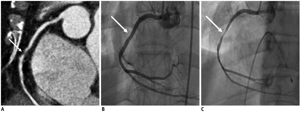
Fig. 2 55-year-old woman with acute chest pain. Curved multiplanar multidetector CT coronary angiography image (A) demonstrating significant discrete stenosis with internal low attenuated area at middle segment of right coronary artery (arrow). Insignificant stenosis is shown at corresponding segment (arrow) on invasive coronary angiography (B). Provoked severe stenosis is revealed at same site (arrow) on invasive coronary angiography after intracoronary administration of ergonovine (C).
Cited by 2 articles
-
Coronary Computed Tomography Angiography for the Diagnosis of Vasospastic Angina: Comparison with Invasive Coronary Angiography and Ergonovine Provocation Test
Jiesuck Park, Hyung-Kwan Kim, Eun-Ah Park, Jun-Bean Park, Seung-Pyo Lee, Whal Lee, Yong-Jin Kim, Dae-Won Sohn
Korean J Radiol. 2019;20(5):719-728. doi: 10.3348/kjr.2018.0847.Comparison of the radiation dose between dual-acquisition coronary computed tomography angiography and coronary angiography for coronary spasm
Soo-Jin Kim, Moo Hyun Kim, Eun-Ju Kang
Kosin Med J. 2022;37(1):46-51. doi: 10.7180/kmj.21.035.
Reference
-
1. Prinzmetal M, Kennamer R, Merliss R, Wada T, Bor N. Angina pectoris. I. A variant form of angina pectoris; preliminary report. Am J Med. 1959. 27:375–388.2. Yasue H, Nakagawa H, Itoh T, Harada E, Mizuno Y. Coronary artery spasm--clinical features, diagnosis, pathogenesis, and treatment. J Cardiol. 2008. 51:2–17.3. Hong MK, Park SW, Lee CW, Ko JY, Kang DH, Song JK, et al. Intravascular ultrasound findings of negative arterial remodeling at sites of focal coronary spasm in patients with vasospastic angina. Am Heart J. 2000. 140:395–401.4. Hong YJ, Jeong MH, Choi YH, Ma EH, Ko JS, Lee MG, et al. Plaque components at coronary sites with focal spasm in patients with variant angina: virtual histology-intravascular ultrasound analysis. Int J Cardiol. 2010. 144:367–372.5. Mollet NR, Cademartiri F, Nieman K, Saia F, Lemos PA, McFadden EP, et al. Multislice spiral computed tomography coronary angiography in patients with stable angina pectoris. J Am Coll Cardiol. 2004. 43:2265–2270.6. Nieman K, Cademartiri F, Lemos PA, Raaijmakers R, Pattynama PM, de Feyter PJ. Reliable noninvasive coronary angiography with fast submillimeter multislice spiral computed tomography. Circulation. 2002. 106:2051–2054.7. Ropers D, Baum U, Pohle K, Anders K, Ulzheimer S, Ohnesorge B, et al. Detection of coronary artery stenoses with thinslice multi-detector row spiral computed tomography and multiplanar reconstruction. Circulation. 2003. 107:664–666.8. Achenbach S, Giesler T, Ropers D, Ulzheimer S, Anders K, Wenkel E, et al. Comparison of image quality in contrastenhanced coronary-artery visualization by electron beam tomography and retrospectively electrocardiogram-gated multislice spiral computed tomography. Invest Radiol. 2003. 38:119–128.9. Achenbach S, Moselewski F, Ropers D, Ferencik M, Hoffmann U, MacNeill B, et al. Detection of calcified and noncalcified coronary atherosclerotic plaque by contrast-enhanced, submillimeter multidetector spiral computed tomography: a segment-based comparison with intravascular ultrasound. Circulation. 2004. 109:14–17.10. Leber AW, Knez A, Becker A, Becker C, von Ziegler F, Nikolaou K, et al. Accuracy of multidetector spiral computed tomography in identifying and differentiating the composition of coronary atherosclerotic plaques: a comparative study with intracoronary ultrasound. J Am Coll Cardiol. 2004. 43:1241–1247.11. Dorgelo J, Willems TP, Geluk CA, van Ooijen PM, Zijlstra F, Oudkerk M. Multidetector computed tomography-guided treatment strategy in patients with non-ST elevation acute coronary syndromes: a pilot study. Eur Radiol. 2005. 15:708–713.12. Ghersin E, Litmanovich D, Dragu R, Rispler S, Lessick J, Ofer A, et al. 16-MDCT coronary angiography versus invasive coronary angiography in acute chest pain syndrome: a blinded prospective study. AJR Am J Roentgenol. 2006. 186:177–184.13. White CS, Kuo D, Kelemen M, Jain V, Musk A, Zaidi E, et al. Chest pain evaluation in the emergency department: can MDCT provide a comprehensive evaluation? AJR Am J Roentgenol. 2005. 185:533–540.14. Leber AW, Knez A, White CW, Becker A, von Ziegler F, Muehling O, et al. Composition of coronary atherosclerotic plaques in patients with acute myocardial infarction and stable angina pectoris determined by contrast-enhanced multislice computed tomography. Am J Cardiol. 2003. 91:714–718.15. von Erffa J, Ropers D, Pflederer T, Schmid M, Marwan M, Daniel WG, et al. Differentiation of total occlusion and high-grade stenosis in coronary CT angiography. Eur Radiol. 2008. 18:2770–2775.16. Hackett D, Larkin S, Chierchia S, Davies G, Kaski JC, Maseri A. Induction of coronary artery spasm by a direct local action of ergonovine. Circulation. 1987. 75:577–582.17. Morikawa Y, Uemura S, Ishigami K, Soeda T, Okayama S, Takemoto Y, et al. Morphological features of coronary arteries in patients with coronary spastic angina: assessment with intracoronary optical coherence tomography. Int J Cardiol. 2011. 146:334–340.18. Saito S, Yamagishi M, Takayama T, Chiku M, Koyama J, Ito K, et al. Plaque morphology at coronary sites with focal spasm in variant angina: study using intravascular ultrasound. Circ J. 2003. 67:1041–1045.
- Full Text Links
-
- Actions
-
Cited
- CITED
-
- Close
- Share
-
- Similar articles
-
- Exercise-Induced Vasospastic Angina With Prominent Regional Wall Motion Abnormality
- A Single Coronary Artery: Right Coronary Artery Originating From the Distal Left Circumflex Artery
- Diagnostic Significance of ECG Ergonovine Provocation Test in Patients with Vasospastic Angina
- The Vasomotor Tone In Vasospastic Angina
- Left Circumflex Coronary Artery Fistula Connected to the Right Bronchial Artery Associated with Bronchiectasis: Multidetector CT and Coronary Angiography Findings
