Korean J Lab Med.
2010 Oct;30(5):507-510. 10.3343/kjlm.2010.30.5.507.
Virulence Characteristics of Sucrose-fermenting Vibrio vulnificus Strains
- Affiliations
-
- 1Department of Emergency Medicine, Chosun University Medical School, Gwangju, Korea.
- 2Research Center for Resistant Cells, Chosun University Medical School, Gwangju, Korea. shsin@chosun.ac.kr
- 3Department of Microbiology, Chosun University Medical School, Gwangju, Korea.
- KMID: 1022042
- DOI: http://doi.org/10.3343/kjlm.2010.30.5.507
Abstract
- We identified 6 sucrose-fermenting Vibrio vulnificus strains and examined their virulence characteristics. They were all encapsulated, motile, capable of producing toxins and utilizing transferrin-bound iron, cytotoxic to cultured cells, and virulent enough to kill mice. They could be definitely identified only by genetic identification methods such as PCR, and not by conventional culture-based identification methods such as API 20E (bioMerieux, France). These results indicate that it is essential to adopt genetic approaches as early as possible in order to avoid misdiagnosis of such strains, especially in clinical situations.
Keyword
MeSH Terms
Figure
-
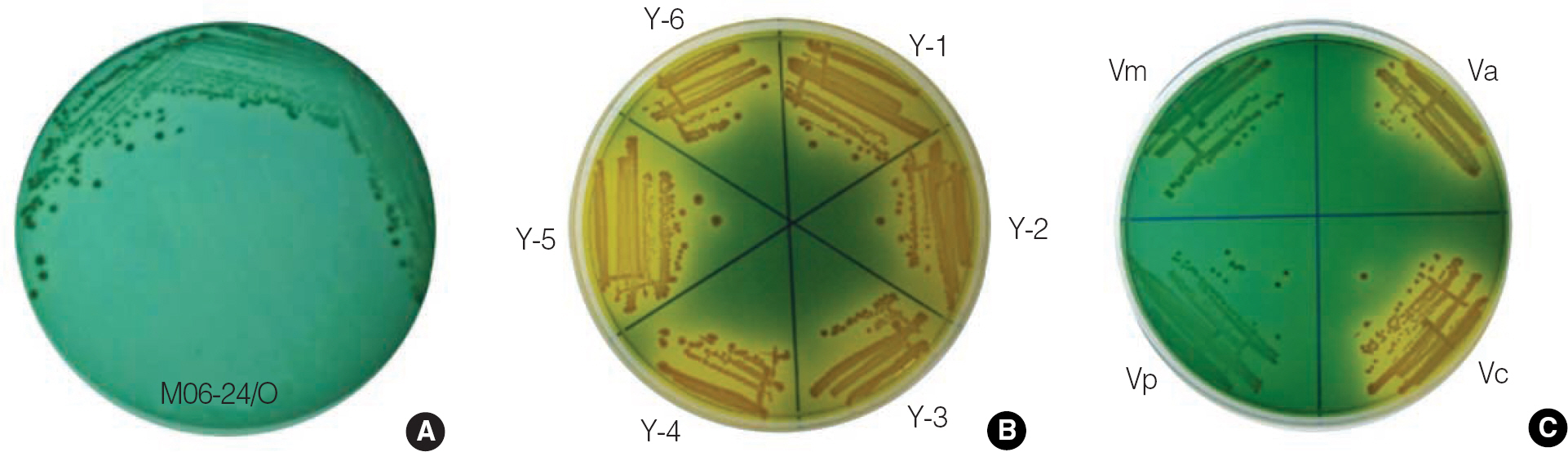
Fig. 1. Sucrose-fermenting Vibrio vulnificus strains forming yellow colonies on thiosulfate citrate bile salts sucrose (TCBS) agar. The M06-24/O strain (A), the 6 sucrose-fermenting V. vulnificus strains (Y-1 to Y-6; B), and other Vibrio species (C), V. alginolyticus ATCC17749 (Va) strain, V. cholerae Non O1 (Vc) strain, V. parahaemolyticus ATCC27519 (Vp) strain, and V. mimicus ATCC33653 (Vm) strain, were used. One colony of each strain grown on Heart Infusion agar was picked, streaked on TCBS agar, and incubated at 37°C for 24 hr.
Reference
-
1.Jones MK., Oliver JD. Vibrio vulnificus: disease and pathogenesis. Infect Immun. 2009. 77:1723–33.2.Harwood VJ., Gandhi JP., Wright AC. Methods for isolation and confirmation of Vibrio vulnificus from oysters and environmental sources: a review. J Microbiol Methods. 2004. 59:301–16.3.Abbott SL., Janda JM., Johnson JA., Farmer III JJ. Vibrio and related organisms. Murray PR, Baron EJ, editors. Manual of clinical microbiology. 9th ed.Washington, DC: American Society of Microbiology;2007. p. 723–33.4.Nagao M., Shimizu Y., Kawada Y., Baba H., Yamada K., Torii K, et al. Two cases of sucrose-fermenting Vibrio vulnificus infection in which 16S rDNA sequencing was useful for diagnosis. Jpn J Infect Dis. 2006. 59:108–10.5.Reddy GP., Hayat U., Abeygunawardana C., Fox C., Wright AC., Maneval DR Jr, et al. Purification and determination of the structure of capsular polysaccharide of Vibrio vulnificus M06-24. J Bacteriol. 1992. 174:2620–30.6.Dalsgaard A., Dalsgaard I., H⊘i L., Larsen JL. Comparison of a commercial biochemical kit and an oligonucleotide probe for identification of environmental isolates of Vibrio vulnificus. Lett Appl Microbiol. 1996. 22:184–8.7.Hill WE., Keasler SP., Trucksess MW., Feng P., Kaysner CA., Lampel KA. Polymerase chain reaction identification of Vibrio vulnificus in artificially contaminated oysters. Appl Environ Microbiol. 1991. 57:707–11.8.Shao CP., Hor LI. Metalloprotease is not essential for Vibrio vulnificus virulence in mice. Infect Immun. 2000. 68:3569–73.9.Kim YR., Lee SE., Kook H., Yeom JA., Na HS., Kim SY, et al. Vibrio vulnificus RTX toxin kills host cells only after contact of the bacteria with host cells. Cell Microbiol. 2008. 10:848–62.10.Kim CM., Park RY., Chun HJ., Kim SY., Rhee JH., Shin SH. Vibrio vulnificus metalloprotease VvpE is essentially required for swarming. FEMS Microbiol Lett. 2007. 269:170–9.11.Kim CM., Park RY., Choi MH., Sun HY., Shin SH. Ferrophilic characteristics of Vibrio vulnificus and potential usefulness of iron chelation therapy. J Infect Dis. 2007. 195:90–8.12.Kataoka E., Honma M., Ohnishi K., Sofuni T., Mizusawa H. Application of highly polymorphic DNA markers to the identification of HeLa cell sublines. In Vitro Cell Dev Biol. 1992. 28A:553–6.
Article
- Full Text Links
-
- Actions
-
Cited
- CITED
-
- Close
- Share
-
- Similar articles
-
- Quorum Sensing System and Virulence Regulation in Vibrio vulnificus
- Comparison of Biological and Genetic Characteristics Between Sucrose-Fermenting and Sucrose-Nonfermenting Vibrio vulnificus Isolates
- Effect of Hydrogen Ion Concentration on the Growth of Vibrio vulnificus
- Cultural Characteristics of Vibrio vulnificus on Various Media
- Biological characteristics and serovars of vibrio vulnificus
