Myositis Ossificans of the Elbow after a Trigger Point Injection
- Affiliations
-
- 1Department of Orthopedic Surgery, Ewha Womans University School of Medicine, Seoul, Korea. sjshin622@ewha.ac.kr
- KMID: 999465
- DOI: http://doi.org/10.4055/cios.2011.3.1.81
Abstract
- Trigger point injection is a simple procedure that is widely performed for relieving pain. Even though there are several complications of trigger point injection, myositis ossificans has not been documented as one of its complications. We treated a patient who suffered from painful limitation of elbow motion and this was caused by myositis ossificans between the insertions of brachialis and supinator muscles after a trigger point injection containing lidocaine mixed with saline, and we also review the relevant medical literature.
MeSH Terms
Figure
-
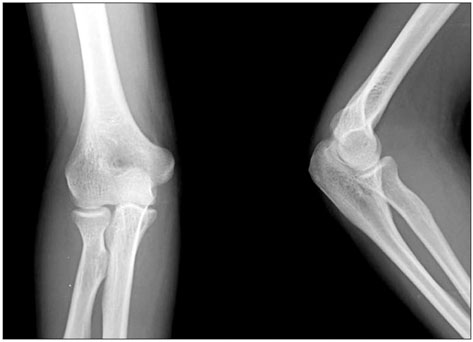
Fig. 1 The preoperative anterior/posterior and lateral radiographs showed soft tissue swelling without a bony lesion.
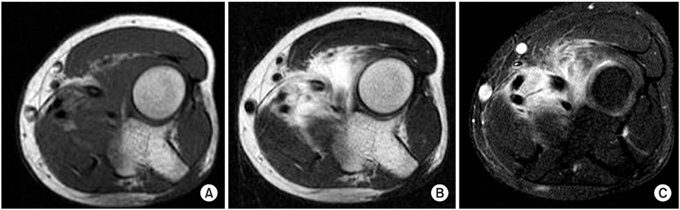
Fig. 2 Magnetic resonance imaging noted a suspicious lesion between the insertion of the brachialis and supinator muscles, and this lesion was regarded as inflammatory tissue without formation of an abscess pocket. This lesion showed (A) low signal intensity on the T1-weighted axial image, (B) high signal intensity on the T2-weighted axial image and (C) a strongly enhanced T1-weighted axial image by gadolinium-DTPA.
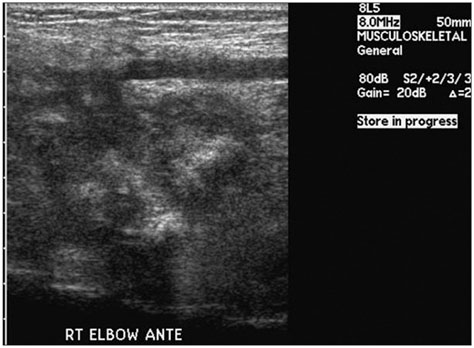
Fig. 3 Ultrasonography demonstrated a hypoechoic area with irregular margins and posterior shadowing.
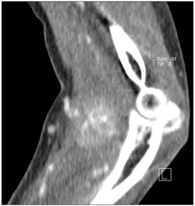
Fig. 4 Computed tomography demonstrated a 4 × 4.6 cm sized high-density mass lesion between the insertion of the brachialis and supinator muscles, and the mass lesion was enhanced at 24 Hounsfield units.
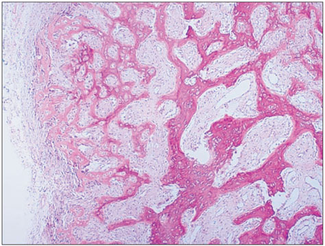
Fig. 5 Pathology demonstrated the extensive osteoid formation rimmed by osteoblasts and a few inflammatory cells and the zoning phenomenon (H&E, ×100).
Reference
-
1. Cheng J, Abdi S. Complications of joint, tendon, and muscle injections. Tech Reg Anesth Pain Manag. 2007. 11(3):141–147.
Article2. Thompson HC 3rd, Garcia A. Myositis ossificans: aftermath of elbow injuries. Clin Orthop Relat Res. 1967. 50:129–134.3. Kaminsky SL, Corcoran D, Chubb WF, Pulla RJ. Myositis ossificans: pedal manifestations. J Foot Surg. 1992. 31(2):173–181.4. Gunduz B, Erhan B, Demir Y. Subcutaneous injections as a risk factor of myositis ossificans traumatica in spinal cord injury. Int J Rehabil Res. 2007. 30(1):87–90.
Article5. Schroder H, Schulz M, Aeissen K. Muscular calcification following injection of vitamin E in newborn infants. Eur J Pediatr. 1984. 142(2):145–146.
Article6. Shirkhoda A, Armin AR, Bis KG, Makris J, Irwin RB, Shetty AN. MR imaging of myositis ossificans: variable patterns at different stages. J Magn Reson Imaging. 1995. 5(3):287–292.
Article7. De Smet AA, Norris MA, Fisher DR. Magnetic resonance imaging of myositis ossificans: analysis of seven cases. Skeletal Radiol. 1992. 21(8):503–507.
Article8. Wieder DL. Treatment of traumatic myositis ossificans with acetic acid iontophoresis. Phys Ther. 1992. 72(2):133–137.
Article9. Kim JH, Chu FC, Woodard HQ, Melamed MR, Huvos A, Cantin J. Radiation-induced soft-tissue and bone sarcoma. Radiology. 1978. 129(2):501–508.
Article10. Viola RW, Hastings H 2nd. Treatment of ectopic ossification about the elbow. Clin Orthop Relat Res. 2000. (370):65–86.
Article
- Full Text Links
-
- Actions
-
Cited
- CITED
-
- Close
- Share
-
- Similar articles
-
- Myositis ossificans progressiva
- Myositis Ossificans Traumatica Causing Ankylosis of the Elbow
- Surgical Treatment of the Myositis Ossificans in Supracondylar Fracture of the Humerus in Children: A Case Report
- Myositis Ossificans in the Finger: A Case Report
- Myositis Ossificans in Rectus Abdominis Muscle: Case Report
