Ultrasonography and the Ultrasound-Based Management of Thyroid Nodules: Consensus Statement and Recommendations
- Affiliations
-
- 1Department of Radiology, Konkuk University Medical Center, Konkuk University School of Medicine, Seoul 143-914, Korea. mdmoonwj@naver.com
- 2Department of Radiology and Research Institute of Radiology, Asan Medical Center, University of Ulsan College of Medicine, Seoul 138-736, Korea.
- 3Department of Radiology, Seoul St. Mary's Hospital, The Catholic University of Korea, Seoul 137-701, Korea.
- 4Department of Radiology, Busan Paik Hospital, Inje University College of Medicine, Busan 614-735, Korea.
- 5Department of Radiology, Research Institute of Radiological Science, Yonsei University College of Medicine, Seoul 120-752, Korea.
- 6Department of Radiology, Dong-A University Medical Center, Dong-A University College of Medicine, Busan 602-715, Korea.
- 7Department of Radiology, Ansan Hospital, Korea University School of Medicine, Gyeonggi-do 425-707, Korea.
- 8Human Medical Imaging & Intervention Center, Seoul 137-902, Korea.
- 9Healthcare System Gangnam Center, Seoul National University Hospital, Seoul 135-984, Korea.
- 10Department of Radiology, Hanyang University Hospital, Hanyang University College of Medicine, Seoul 133-792, Korea.
- 11Department of Radiology, SMG-SNU Boramae Medical Center, Seoul National University College of Medicine, Seoul 156-707, Korea.
- KMID: 991677
- DOI: http://doi.org/10.3348/kjr.2011.12.1.1
Abstract
- The detection of thyroid nodules has become more common with the widespread use of ultrasonography (US). US is the mainstay for detecting and making the differential diagnosis of thyroid nodules as well as for providing guidance for a biopsy. The Task Force on Thyroid Nodules of the Korean Society of Thyroid Radiology has developed recommendations for the US diagnosis and US-based management of thyroid nodules. The review and recommendations in this report have been based on a comprehensive analysis of the current literature, the results of multicenter studies and from the consensus of experts.
MeSH Terms
Figure
-
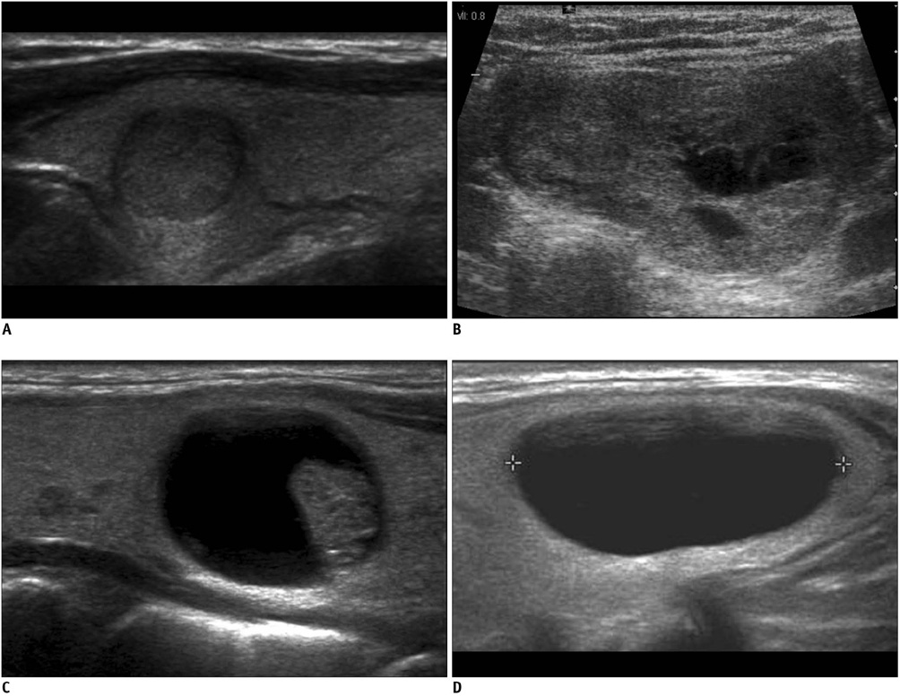
Fig. 1 Internal content of thyroid nodules. A. Solid B. Predominantly solid C. Predominantly cystic D. Cystic

Fig. 2 US findings of spongiform appearance are shown Transverse US image of benign nodular hyperplasia shows well-defined smooth isoechoic mass with a spongiform appearance (arrows).

Fig. 3 Shape of thyroid nodules. Corresponding schematic drawings are shown in upper panel. A. Ovoid-to-round shape B. Taller-than-wide shape C. Irregular shape

Fig. 4 Margin of thyroid nodules. A. Smooth margin B. Spiculated or microlobulated margin (arrow) C. Ill-defined margin (arrows)
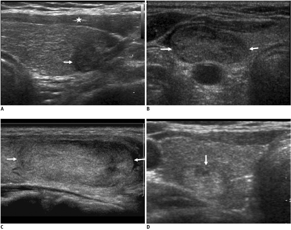
Fig. 5 Echogenicity of thyroid nodules. A. Marked hypoechogenicity of nodule is shown. Note more hypoechoic nature of nodule (arrow) as compared to that of strap muscles (asterisk). B. Hypoechogenicity of nodule (arrows) C. Isoechogenicity of nodule (arrows) D. Hyperechogenicity of nodule (arrow)
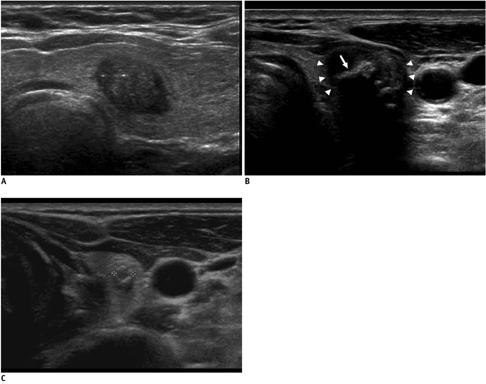
Fig. 6 Calcifications of thyroid nodules. A. Microcalcification within nodule as echogenic focus B. Macrocalcification (arrow) in center of nodule (white triangles) C. Rim calcification in small nodule (calipers).
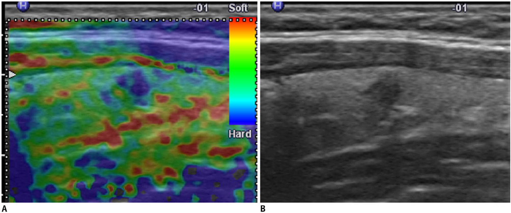
Fig. 7 US elastography of thyroid nodules. A. Elastography shows nodule with hard consistency as blue relative to green background. B. Longitudinal US image shows same nodule with suspicious malignant US features. Nodule was proven to be papillary carcinoma.

Fig. 8 Flowchart for strategy for follow-up US and US-guided fine needle aspiration (USFNA) biopsy according to US findings and cytology results of thyroid nodules. Dotted arrow means that surgery is not strongly recommended, but it can be considered according to individual situations. AUS = atypia of undetermined significance, FLUS = follicular lesion of undetermined significance.
Cited by 12 articles
-
Feasibility of Adjustable Electrodes for Radiofrequency Ablation of Benign Thyroid Nodules
Jiyeong Lee, Jung Hee Shin, Soo Yeon Hahn, Ko Woon Park, Ji Soo Choi
Korean J Radiol. 2020;21(3):377-383. doi: 10.3348/kjr.2019.0724.Mixed Echoic Thyroid Nodules on Ultrasound: Approach to Management
Yu-Mee Sohn, Jung Hyun Yoon, Hee Jung Moon, Eun-Kyung Kim, Jin Young Kwak
Yonsei Med J. 2012;53(4):812-819. doi: 10.3349/ymj.2012.53.4.812.Diagnostic Role of Conventional Ultrasonography and Shearwave Elastography in Asymptomatic Patients with Diffuse Thyroid Disease: Initial Experience with 57 Patients
Injoong Kim, Eun-Kyung Kim, Jung Hyun Yoon, Kyung Hwa Han, Eun Ju Son, Hee Jung Moon, Jin Young Kwak
Yonsei Med J. 2014;55(1):247-253. doi: 10.3349/ymj.2014.55.1.247.Clinical Characteristics and Incidence of Thyroid Nodule in the Male Population for Health Check-up
Yeon Jin Jeon, Young Teag Koh, Seung Jong Oh, Min Young Koo
Korean J Endocr Surg. 2015;15(4):93-98. doi: 10.16956/kjes.2015.15.4.93.Thyroid Ultrasonography: Pitfalls and Techniques
Seon Hyeong Choi, Eun-Kyung Kim, Soo Jin Kim, Jin Young Kwak
Korean J Radiol. 2014;15(2):267-276. doi: 10.3348/kjr.2014.15.2.267.Primary Imaging Test and Appropriate Biopsy Methods for Thyroid Nodules: Guidelines by Korean Society of Radiology and National Evidence-Based Healthcare Collaborating Agency
Eun Ju Ha, Hyun Kyung Lim, Jung Hyun Yoon, Jung Hwan Baek, Kyung Hyun Do, Miyoung Choi, Jin A Choi, Min Lee, Dong Gyu Na,
Korean J Radiol. 2018;19(4):623-631. doi: 10.3348/kjr.2018.19.4.623.Evaluation of Modified Core-Needle Biopsy in the Diagnosis of Thyroid Nodules
Soomin Ahn, Sejin Jung, Ji-Ye Kim, Jung Hee Shin, Soo Yeon Hahn, Young Lyun Oh
Korean J Radiol. 2018;19(4):656-664. doi: 10.3348/kjr.2018.19.4.656.History of Korean Society of Thyroid Radiology
Dong Gyu Na, Jung Hwan Baek
Int J Thyroidol. 2018;11(1):11-14. doi: 10.11106/ijt.2018.11.1.11.Guidelines for Primary Imaging Test and Biopsy Methods in the Diagnosis of Thyroid Nodules: Joint Report by the Korean Society of Radiology and National Evidence-Based Healthcare Collaborating Agency
Hyun Kyung Lim, Eun Ju Ha, In Young Youn, Jung Hyun Yoon, Jung Hwan Baek, Kyung Hyun Do, Miyoung Choi, Jin A Choi, Min Lee, Dong Gyu Na
J Korean Soc Radiol. 2018;79(1):1-10. doi: 10.3348/jksr.2018.79.1.1.Current Guidelines for Fine Needle Aspiration of Thyroid Nodules
Jeonghun Lee, Euy Young Soh
Korean J Endocr Surg. 2015;15(1):1-5. doi: 10.16956/kjes.2015.15.1.1.Ultrasonography Diagnosis of Thyroid Nodules and Cervical Metastatic Lymph Nodes
Dong Gyu Na, Young Hen Lee
Int J Thyroidol. 2016;9(1):1-8. doi: 10.11106/ijt.2016.9.1.1.Evaluation of Diagnostic Performance of Screening Thyroid Ultrasonography and Imaging Findings of Screening-Detected Thyroid Cancer
Jeongin Yoo, Hye Shin Ahn, Soo Jin Kim, Sung Hee Park, Mirinae Seo, Semin Chong
Cancer Res Treat. 2018;50(1):11-18. doi: 10.4143/crt.2016.600.
Reference
-
1. Harach HR, Franssila KO, Wasenius VM. Occult papillary carcinoma of the thyroid. A "normal" finding in Finland. A systematic autopsy study. Cancer. 1985. 56:531–538.2. Brander A, Viikinkoski P, Nickels J, Kivisaari L. Thyroid gland: US screening in a random adult population. Radiology. 1991. 181:683–687.3. Tan GH, Gharib H. Thyroid incidentalomas: management approaches to nonpalpable nodules discovered incidentally on thyroid imaging. Ann Intern Med. 1997. 126:226–231.4. Frates MC, Benson CB, Charboneau JW, Cibas ES, Clark OH, Coleman BG, et al. Management of thyroid nodules detected at US: Society of Radiologists in Ultrasound consensus conference statement. Radiology. 2005. 237:794–800.5. Nam-Goong IS, Kim HY, Gong G, Lee HK, Hong SJ, Kim WB, et al. Ultrasonography-guided fine-needle aspiration of thyroid incidentaloma: correlation with pathological findings. Clin Endocrinol (Oxf). 2004. 60:21–28.6. 2005 annual report of the Korea central cancer registry [www document]. National Cancer Information Center K. last accessed; Oct 2008. Available at : http://www.cancer.go.kr.7. Gharib H, Papini E, Valcavi R, Baskin HJ, Crescenzi A, Dottorini ME, et al. American Association of Clinical Endocrinologists and Associazione Medici Endocrinologi medical guidelines for clinical practice for the diagnosis and management of thyroid nodules. Endocr Pract. 2006. 12:63–102.8. American Thyroid Association (ATA) Guidelines Taskforce on Thyroid Nodules and Differentiated Thyroid Cancer. Cooper DS, Doherty GM, Haugen BR, Kloos RT, Lee SL, et al. Revised American Thyroid Association management guidelines for patients with thyroid nodules and differentiated thyroid cancer. Thyroid. 2009. 19:1167–1214.9. Moon WJ, Jung SL, Lee JH, Na DG, Baek JH, Lee YH, et al. Benign and malignant thyroid nodules: US differentiation--multicenter retrospective study. Radiology. 2008. 247:762–770.10. Moon WJ, Na DG, Jung SL, Lee JH, Kim J, Kim HS, et al. Recommendations for ultrasound-based management of thyroid nodules. 62nd Scientific Assembly of the Korean Radiological Society. 2006. Seoul: The Korean Radiological Society.11. Brander AE, Viikinkoski VP, Nickels JI, Kivisaari LM. Importance of thyroid abnormalities detected at US screening: a 5-year follow-up. Radiology. 2000. 215:801–806.12. Kuma K, Matsuzuka F, Yokozawa T, Miyauchi A, Sugawara M. Fate of untreated benign thyroid nodules: results of long-term follow-up. World J Surg. 1994. 18:495–498.13. Alexander EK, Hurwitz S, Heering JP, Benson CB, Frates MC, Doubilet PM, et al. Natural history of benign solid and cystic thyroid nodules. Ann Intern Med. 2003. 138:315–318.14. Hoang JK, Lee WK, Lee M, Johnson D, Farrell S. US features of thyroid malignancy: pearls and pitfalls. Radiographics. 2007. 27:847–860.15. Papini E, Petrucci L, Guglielmi R, Panunzi C, Rinaldi R, Bacci V, et al. Long-term changes in nodular goiter: a 5-year prospective randomized trial of levothyroxine suppressive therapy for benign cold thyroid nodules. J Clin Endocrinol Metab. 1998. 83:780–783.16. Brauer VF, Eder P, Miehle K, Wiesner TD, Hasenclever H, Paschke R. Interobserver variation for ultrasound determination of thyroid nodule volumes. Thyroid. 2005. 15:1169–1175.17. Chan BK, Desser TS, McDougall IR, Weigel RJ, Jeffrey RB Jr. Common and uncommon sonographic features of papillary thyroid carcinoma. J Ultrasound Med. 2003. 22:1083–1090.18. Watters DA, Ahuja AT, Evans RM, Chick W, King WW, Metreweli C, et al. Role of ultrasound in the management of thyroid nodules. Am J Surg. 1992. 164:654–657.19. Lee MJ, Kim EK, Kwak JY, Kim MJ. Partially cystic thyroid nodules on ultrasound: probability of malignancy and sonographic differentiation. Thyroid. 2009. 19:341–346.20. Hatabu H, Kasagi K, Yamamoto K, Iida Y, Misaki T, Hidaka A, et al. Cystic papillary carcinoma of the thyroid gland: a new sonographic sign. Clin Radiol. 1991. 43:121–124.21. Bonavita JA, Mayo J, Babb J, Bennett G, Oweity T, Macari M, et al. Pattern recognition of benign nodules at ultrasound of the thyroid: which nodules can be left alone? AJR Am J Roentgenol. 2009. 193:207–213.22. Moon WJ, Kwag HJ, Na DG. Are there any specific ultrasound findings of nodular hyperplasia ("leave me alone" lesion) to differentiate it from follicular adenoma? Acta Radiol. 2009. 50:383–388.23. Kim EK, Park CS, Chung WY, Oh KK, Kim DI, Lee JT, et al. New sonographic criteria for recommending fine-needle aspiration biopsy of nonpalpable solid nodules of the thyroid. AJR Am J Roentgenol. 2002. 178:687–691.24. Alexander EK, Marqusee E, Orcutt J, Benson CB, Frates MC, Doubilet PM, et al. Thyroid nodule shape and prediction of malignancy. Thyroid. 2004. 14:953–958.25. Stavros AT, Thickman D, Rapp CL, Dennis MA, Parker SH, Sisney GA. Solid breast nodules: use of sonography to distinguish between benign and malignant lesions. Radiology. 1995. 196:123–134.26. Papini E, Guglielmi R, Bianchini A, Crescenzi A, Taccogna S, Nardi F, et al. Risk of malignancy in nonpalpable thyroid nodules: predictive value of ultrasound and color-Doppler features. J Clin Endocrinol Metab. 2002. 87:1941–1946.27. Reading CC, Charboneau JW, Hay ID, Sebo TJ. Sonography of thyroid nodules: a "classic pattern" diagnostic approach. Ultrasound Q. 2005. 21:157–165.28. Khoo ML, Asa SL, Witterick IJ, Freeman JL. Thyroid calcification and its association with thyroid carcinoma. Head Neck. 2002. 24:651–655.29. Peccin S, de Castsro JA, Furlanetto TW, Furtado AP, Brasil BA, Czepielewski MA. Ultrasonography: is it useful in the diagnosis of cancer in thyroid nodules? J Endocrinol Invest. 2002. 25:39–43.30. Kwak MS, Baek JH, Kim YS, Jeong HJ. Patterns and significance of peripheral calcifications of thyroid tumors seen on ultrasound. J Korean Radiol Soc. 2005. 53:401–405.31. Yoon DY, Lee JW, Chang SK, Choi CS, Yun EJ, Seo YL, et al. Peripheral calcification in thyroid nodules: ultrasonographic features and prediction of malignancy. J Ultrasound Med. 2007. 26:1349–1355.32. Kim BM, Kim MJ, Kim EK, Kwak JY, Hong SW, Son EJ, et al. Sonographic differentiation of thyroid nodules with eggshell calcifications. J Ultrasound Med. 2008. 27:1425–1430.33. Propper RA, Skolnick ML, Weinstein BJ, Dekker A. The nonspecificity of the thyroid halo sign. J Clin Ultrasound. 1980. 8:129–132.34. Lu C, Chang TC, Hsiao YL, Kuo MS. Ultrasonographic findings of papillary thyroid carcinoma and their relation to pathologic changes. J Formos Med Assoc. 1994. 93:933–938.35. Tamsel S, Demirpolat G, Erdogan M, Nart D, Karadeniz M, Uluer H, et al. Power Doppler US patterns of vascularity and spectral Doppler US parameters in predicting malignancy in thyroid nodules. Clin Radiol. 2007. 62:245–251.36. Moon HJ, Kwak JY, Kim MJ, Son EJ, Kim EK. Can vascularity at power Doppler US help predict thyroid malignancy? Radiology. 2010. 255:260–269.37. Lyshchik A, Higashi T, Asato R, Tanaka S, Ito J, Mai JJ, et al. Thyroid gland tumor diagnosis at US elastography. Radiology. 2005. 237:202–211.38. Kim SJ, Kim EK, Park CS, Chung WY, Oh KK, Yoo HS. Ultrasound-guided fine-needle aspiration biopsy in nonpalpable thyroid nodules: is it useful in infracentimetric nodules? Yonsei Med J. 2003. 44:635–640.39. Wienke JR, Chong WK, Fielding JR, Zou KH, Mittelstaedt CA. Sonographic features of benign thyroid nodules: interobserver reliability and overlap with malignancy. J Ultrasound Med. 2003. 22:1027–1031.40. Iannuccilli JD, Cronan JJ, Monchik JM. Risk for malignancy of thyroid nodules as assessed by sonographic criteria: the need for biopsy. J Ultrasound Med. 2004. 23:1455–1464.41. Frates MC, Benson CB, Doubilet PM, Cibas ES, Marqusee E. Can color Doppler sonography aid in the prediction of malignancy of thyroid nodules? J Ultrasound Med. 2003. 22:127–131.42. Popowicz B, Klencki M, Lewiński A, Słowińska-Klencka D. The usefulness of sonographic features in selection of thyroid nodules for biopsy in relation to the nodule's size. Eur J Endocrinol. 2009. 161:103–111.43. Ahuja A, Chick W, King W, Metreweli C. Clinical significance of the comet-tail artifact in thyroid ultrasound. J Clin Ultrasound. 1996. 24:129–133.44. Kim SH, Kim BS, Jung SL, Lee JW, Yang PS, Kang BJ, et al. Ultrasonographic findings of medullary thyroid carcinoma: a comparison with papillary thyroid carcinoma. Korean J Radiol. 2009. 10:101–105.45. Jeh SK, Jung SL, Kim BS, Lee YS. Evaluating the degree of conformity of papillary carcinoma and follicular carcinoma to the reported ultrasonographic findings of malignant thyroid tumor. Korean J Radiol. 2007. 8:192–197.46. Kim DS, Kim JH, Na DG, Park SH, Kim E, Chang KH, et al. Sonographic features of follicular variant papillary thyroid carcinomas in comparison with conventional papillary thyroid carcinomas. J Ultrasound Med. 2009. 28:1685–1692.47. Mazzaferri EL. Management of a solitary thyroid nodule. N Engl J Med. 1993. 328:553–559.48. Mazzaferri EL, Jhiang SM. Long-term impact of initial surgical and medical therapy on papillary and follicular thyroid cancer. Am J Med. 1994. 97:418–428.49. Pazaitou-Panayiotou K, Capezzone M, Pacini F. Clinical features and therapeutic implication of papillary thyroid microcarcinoma. Thyroid. 2007. 17:1085–1092.50. Baudin E, Travagli JP, Ropers J, Mancusi F, Bruno-Bossio G, Caillou B, et al. Microcarcinoma of the thyroid gland: the Gustave-Roussy Institute experience. Cancer. 1998. 83:553–559.51. Chow SM, Law SC, Au SK, Mang O, Yau S, Yuen KT, et al. Changes in clinical presentation, management and outcome in 1348 patients with differentiated thyroid carcinoma: experience in a single institute in Hong Kong, 1960-2000. Clin Oncol (R Coll Radiol). 2003. 15:329–336.52. Ito Y, Uruno T, Nakano K, Takamura Y, Miya A, Kobayashi K, et al. An observation trial without surgical treatment in patients with papillary microcarcinoma of the thyroid. Thyroid. 2003. 13:381–387.53. Noguchi S, Yamashita H, Murakami N, Nakayama I, Toda M, Kawamoto H. Small carcinomas of the thyroid. A long-term follow-up of 867 patients. Arch Surg. 1996. 131:187–191.54. Noguchi S, Yamashita H, Uchino S, Watanabe S. Papillary microcarcinoma. World J Surg. 2008. 32:747–753.55. Kim DW, Lee EJ, Kim SH, Kim TH, Lee SH, Kim DH, et al. Ultrasound-guided fine-needle aspiration biopsy of thyroid nodules: comparison in efficacy according to nodule size. Thyroid. 2009. 19:27–31.56. Mazzaferri EL, Sipos J. Should all patients with subcentimeter thyroid nodules undergo fine-needle aspiration biopsy and preoperative neck ultrasonography to define the extent of tumor invasion? Thyroid. 2008. 18:597–602.57. Asanuma K, Kobayashi S, Shingu K, Hama Y, Yokoyama S, Fujimori M, et al. The rate of tumour growth does not distinguish between malignant and benign thyroid nodules. Eur J Surg. 2001. 167:102–105.58. Erdogan MF, Gursoy A, Erdogan G. Natural course of benign thyroid nodules in a moderately iodine-deficient area. Clin Endocrinol (Oxf). 2006. 65:767–771.59. Cibas ES, Ali SZ. The Bethesda System for Reporting Thyroid Cytopathology. Thyroid. 2009. 19:1159–1165.60. Alexander EK, Heering JP, Benson CB, Frates MC, Doubilet PM, Cibas ES, et al. Assessment of nondiagnostic ultrasoundguided fine needle aspirations of thyroid nodules. J Clin Endocrinol Metab. 2002. 87:4924–4927.61. Ogawa Y, Kato Y, Ikeda K, Aya M, Ogisawa K, Kitani K, et al. The value of ultrasound-guided fine-needle aspiration cytology for thyroid nodules: an assessment of its diagnostic potential and pitfalls. Surg Today. 2001. 31:97–101.62. Kwak JY, Koo H, Youk JH, Kim MJ, Moon HJ, Son EJ, et al. Value of US correlation of a thyroid nodule with initially benign cytologic results. Radiology. 2010. 254:292–300.63. Black WC, Welch HG. Advances in diagnostic imaging and overestimations of disease prevalence and the benefits of therapy. N Engl J Med. 1993. 328:1237–1243.
- Full Text Links
-
- Actions
-
Cited
- CITED
-
- Close
- Share
-
- Similar articles
-
- 2021 Korean Thyroid Imaging Reporting and Data System (2021-K-TIRADS) and Imaging-Based Management of Thyroid Nodules: Korean Society of Thyroid Radiology Consensus Statement and Recommendations
- Standardized Imaging and Reporting for Thyroid Ultrasound: Korean Society of Thyroid Radiology Consensus Statement and Recommendation
- Clinical applications of Doppler ultrasonography for thyroid disease: consensus statement by the Korean Society of Thyroid Radiology
- Use of the ultrasound-based total malignancy score in the management of thyroid nodules
- Indications for Fine Needle Aspiration in Thyroid Nodules
