Clear Cell Hidradenoma of the Axilla: a Case Report with Literature Review
- Affiliations
-
- 1Department of Radiology, Yonsei University College of Medicine, Seoul 135-720, Korea. ejsonrd@yuhs.ac
- 2Department of Surgery, Yonsei University College of Medicine, Seoul 135-720, Korea.
- KMID: 984906
- DOI: http://doi.org/10.3348/kjr.2010.11.4.490
Abstract
- Clear cell hidradenoma is an uncommon benign skin appendageal tumor that typically involves the dermal layer of the head, face, and extremities. The breast is a rare site for this lesion, with only two documented cases, which were determined based on mammogram and sonogram findings. We present a case of clear cell hidradenoma of the axillary tail with radiological findings and a literature review.
Keyword
MeSH Terms
Figure
-
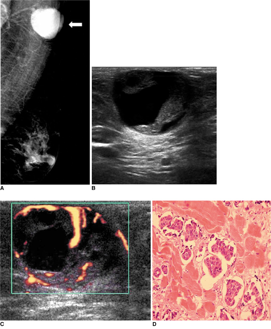
Fig. 1 56-year-old woman with soft, mobile mass in left axillary tail for six months. A. Medio-lateral oblique mammogram showing round, well-circumscribed, high-density mass in left axilla (white arrow). B. Ultrasound showing oval, well-circumscribed, complex mass with posterior acoustic enhancement in subcutaneous fat layer of left axillary tail. C. Power Doppler study revealed increased vascularity in peripheral area and internal solid portion of mass. D. Microscopically, tumor is predominantly composed of clear cells with ductal differentiation and eosinophilic cytoplasm (Hematoxylin & Eosin stain, ×400).
Cited by 1 articles
-
Clear Cell Hidradenoma and Hidradenocarcinoma Arising from Benign Hidradenoma: Imaging Findings of Ultrasonography and CT
Jiyeon Ha, Hye Won Chung, Joon Seon Song
J Korean Soc Radiol. 2019;80(4):768-772. doi: 10.3348/jksr.2019.80.4.768.
Reference
-
1. Jin W, Kim GY, Lew BL, Yang DM, Kim HC, Ryu JK, et al. Sonographic findings of an eccrine spiradenoma: case report and literature review. J Ultrasound Med. 2008. 27:813–818.2. Mullaney PJ, Becker E, Graham B, Ghazarian D, Riddell RH, Salonen DC. Benign hidradenoma: magnetic resonance and ultrasound features of two cases. Skeletal Radiol. 2007. 36:1185–1190.3. Ghai S, Bukhanov K. Eccrine acrospiroma of breast: mammographic and ultrasound findings. Clin Radiol. 2004. 59:1142–1144.4. Ohi Y, Umekita Y, Rai Y, Kukita T, Sagara Y, Sagara Y, et al. Clear cell hidradenoma of the breast: a case report with review of the literature. Breast Cancer. 2007. 14:307–311.5. Shaikh-Naidu N, Breitbart A. Eccrine spiradenoma of the upper extremity: case report and an algorithm for management. Eur J Plast Surg. 2003. 26:160–163.6. Revis P, Chyu J, Medenica M. Multiple eccrine spiradenoma: case report and review. J Cutan Pathol. 1988. 15:226–229.7. El Demellawy D, Daya D, Alowami S. Clear cell hidradenoma: an unusual vulvar tumor. Int J Gynecol Pathol. 2008. 27:457–460.8. Hernández-Pérez E, Cestoni-Parducci R. Nodular hidradenoma and hidradenocarcinoma. A 10-year review. J Am Acad Dermatol. 1985. 12:15–20.9. Han YD, Huan Y, Deng JL, Zhang YG, Zhang CH. MRI appearance of multiple eccrine spiradenoma. Br J Radiol. 2007. 80:E27–E29.
