Estrogen Antagonist and Development of Macular Hole
- Affiliations
-
- 1Department of Ophthalmology, Samsung Medical Center, Sungkyunkwan University School of Medicine, Seoul, Korea. swkang@skku.edu
- 2Doctor Lee's Eye Clinic, Suwon, Korea.
- 3Department of Ophthalmology, Konkuk University School of Medicine, Seoul, Korea.
- KMID: 974326
- DOI: http://doi.org/10.3341/kjo.2010.24.5.306
Abstract
- To describe the clinical and optical coherence tomography (OCT) features of a macular hole (MH) or its precursor lesion in patients treated with systemic antiestrogen agents. We reviewed the medical history of the patient, ophthalmic examination, and both fundus and OCT findings. Three female patients receiving antiestrogen therapy sought treatment for visual disturbance. All of the patients showed foveal cystic changes with outer retinal defect upon OCT. Visual improvement was achieved through surgery for the treatment of MH in two patients. Antiestrogen therapy may result in MH or its precursor lesion, in addition to perifoveal refractile deposits. OCT examination would be helpful for early detection in such cases.
MeSH Terms
Figure
-
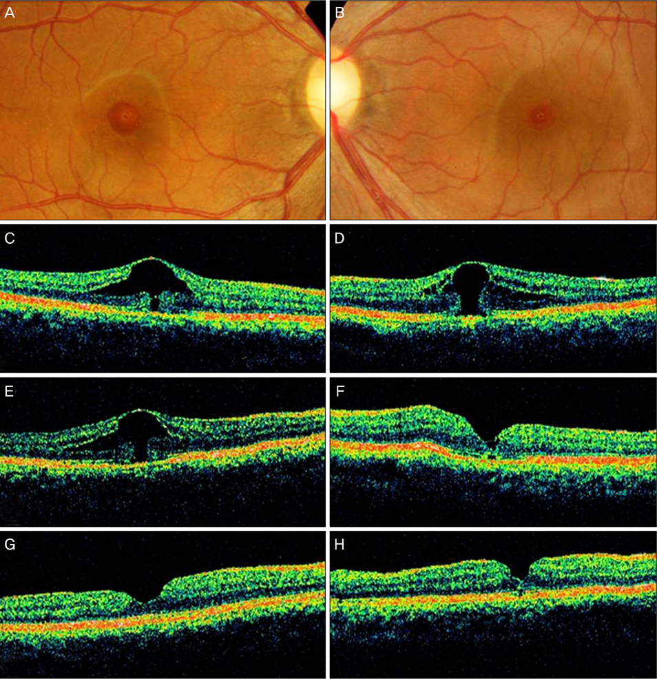
Fig. 1 Fundus photograph and optical coherence tomographic images of both eyes of patient 1. (A,C) At presentation, the image shows foveal cystic changes with outer retinal defect in the patient's right eye. (B,D) At presentation, the image shows a stage 1b macular hole in the left eye. (E) The enlargement of outer foveal defect is noted in the right eye 4 months after initial presentation, with further deterioration of vision. (F) However, improved foveal contour and vision is noted after macular hole surgery in the left eye. (G,H) The closure of macular hole is achieved in both eyes after completing the surgery in both eyes.
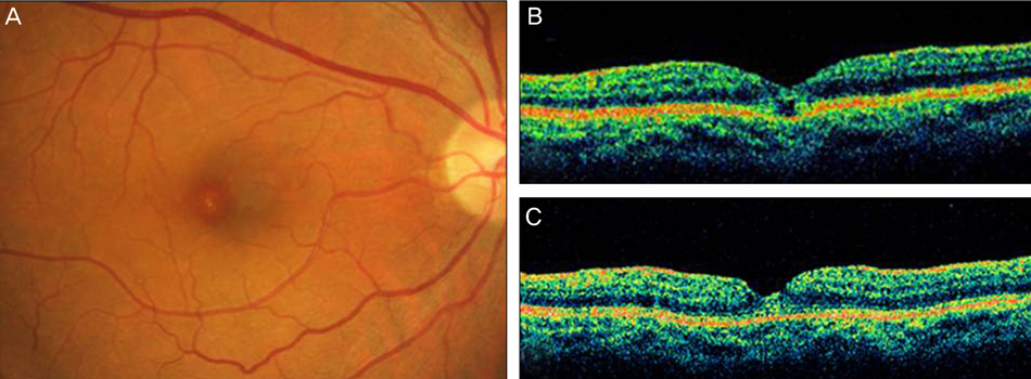
Fig. 2 Right eye of patient 2. Fundus photographic image (A) shows foveal cystic change, and the optical coherence tomographic image (B) shows a focal outer retinal defect at initial presentation. The contour of the fovea returns to normal after macular hole surgery.
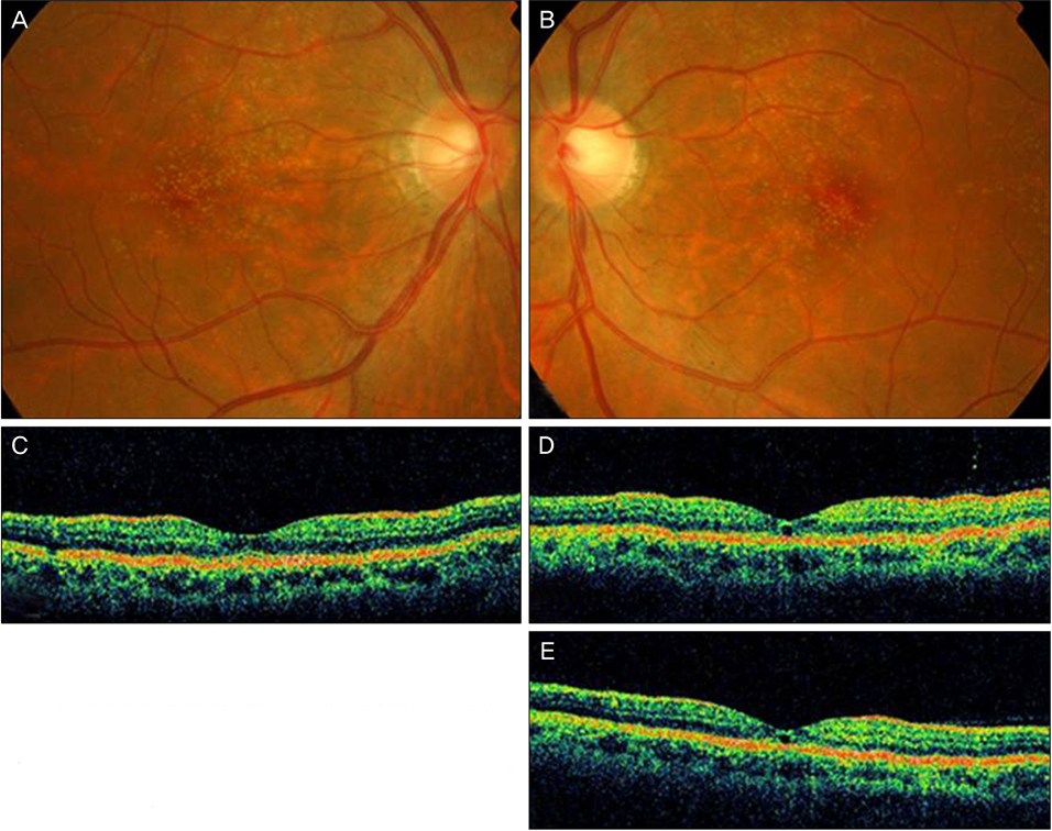
Fig. 3 Fundus photographic images of both eyes in patient 3. At initial presentation, small multiple refractile deposits are noted in both of the patient's eyes (A,B). Optical coherence tomographic (OCT) scan of the macula shows no specific abnormality in the right eye (C); however, a focal defect of the outer retinal layer is noted in the left eye (D). No interval change in OCT findings is noted after 5 months (E).
Cited by 2 articles
-
Uveoretinal Adverse Effects Presented during Systemic Anticancer Chemotherapy: a 10-Year Single Center Experience
Ah Ran Cho, Young Hee Yoon, June-Gone Kim, Yoon Jeon Kim, Joo Yong Lee
J Korean Med Sci. 2018;33(7):. doi: 10.3346/jkms.2018.33.e55.A Case of Postpartum Macular Hole in a Young Woman
Byoung Seon Kim, Byung Jae Kim, Yong Seop Han, Jong Moon Park, In Young Chung
J Korean Ophthalmol Soc. 2015;56(3):463-465. doi: 10.3341/jkos.2015.56.3.463.
Reference
-
1. Bourla DH, Sarraf D, Schwartz SD. Peripheral retinopathy and maculopathy in high-dose tamoxifen therapy. Am J Ophthalmol. 2007. 144:126–128.2. Gualino V, Cohen SY, Delyfer MN, et al. Optical coherence tomography findings in tamoxifen retinopathy. Am J Ophthalmol. 2005. 140:757–758.3. Cronin BG, Lekich CK, Bourke RD. Tamoxifen therapy conveys increased risk of developing a macular hole. Int Ophthalmol. 2005. 26:101–105.4. Bernstein PS, DellaCroce JT. Diagnostic & therapeutic challenges. Tamoxifen toxicity. Retina. 2007. 27:982–988.5. Evans JR, Schwartz SD, McHugh JD, et al. Systemic risk factors for idiopathic macular holes: a case-control study. Eye (Lond). 1998. 12(Pt 2):256–259.6. The Eye Disease Case-Control Study Group. Risk factors for idiopathic macular holes. Am J Ophthalmol. 1994. 118:754–761.
- Full Text Links
-
- Actions
-
Cited
- CITED
-
- Close
- Share
-
- Similar articles
-
- Analysis of Systemic Risk Factors in Idiopathic Macular Hole
- A Case Report of Closed Traumatic Macular Hole after Intravit real Gas Injection
- Eccentric Macular Hole Formation After Macular Hole Surgery
- Influence of the Macular Curvature on Foveal Migration after Macular Hole Surgery
- Spontaneous Disappearance of A Traumatic Macular Hole
