Korean J Ophthalmol.
2010 Apr;24(2):119-122. 10.3341/kjo.2010.24.2.119.
Case Report of Acremonium Intraocular Infection after Cataract Extraction
- Affiliations
-
- 1Department of Ophthalmology, Asan Medical Center, University of Ulsan College of Medicine, Seoul, Korea. yhyoon@amc.seoul.kr
- KMID: 946004
- DOI: http://doi.org/10.3341/kjo.2010.24.2.119
Abstract
- A 64-year-old woman was referred to our clinic for the treatment of chronic uveitis in her left eye, which had started two weeks after an uncomplicated cataract extraction. She was treated with topical steroids with an initially good response, yet she subsequently developed severe inflammation and plaque-like material around the intraocular lens, despite continuous steroid therapy. She underwent pars plana vitrectomy, smear and culture of the aqueous and vitreous fluids, and intravitreal antibiotic injection under the impression of Propionibacterium acne (P. acne) endophthalmitis. As a result of the smear and culture of the vitreous fluid identified as an Acremonium species, she was treated with intravenous amphotericin B injections for five days, followed by oral voriconazole administration. During the post-operative 18-month follow-up, she was stable without significant relapse of uveitis. In this case, the best correction of visual acuity was an improvement from 20/40 to 20/20.
MeSH Terms
Figure
-
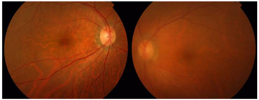
Fig. 1 Fundus photograph at the time of presentation. The left eye is hazy due to vitreous opacity (right).
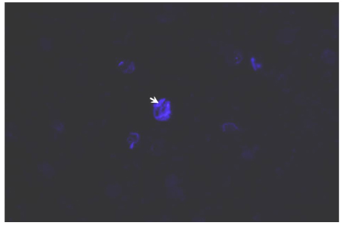
Fig. 2 KOH-Calcofluor staining of vitreous fluid obtained during pars plana vitrectomy H & E, ×400. Septum of fungal hyphae (white arrow).

Fig. 3 Gram staining of the vitreous fluid collected during pars plana vitrectomy, ×1000. WBC (arrow head), septated hyphae (arrow).
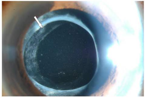
Fig. 4 Anterior segment photograph taken 7 days after surgery. The thin plaque-like material (white arrow) gradually decreased over time.
Reference
-
1. Meredith TA. Ryan SJ, Hinton DR, Schachat AP, Wilkinson CP, editors. Vitrectomy for infectious endophthalmitis. Retina. 2006. Vol. 3:4th ed. Philadelphia: Mosby;2260–2261.2. Scott IU, Flynn HW Jr, Miller D. Delayed-onset endophthalmitis following cataract surgery caused by Acremonium strictum. Ophthalmic Surg Lasers Imaging. 2005. 36:506–507.3. Weissgold DJ, Maguire AM, Brucker AJ. Management of postoperative Acremonium endophthalmitis. Ophthalmology. 1996. 103:749–756.4. Cameron JA, Badawi EM, Hoffman PA, Tabara KF. Chronic endophthalmitis caused by Acremonium falciforme. Can J Ophthalmol. 1996. 31:367–368.5. Fridkin SK, Kremer FB, Bland LA, et al. Acremonium kiliense endophthalmitis that occurred after cataract extraction in an ambulatory surgical care and was traced to an environmental reservoir. Clin Infect Dis. 1996. 22:222–227.6. Fleming RV, Walsh TJ, Anaissie EJ. Emerging and less common fungal pathogens. Infect Dis Clin North Am. 2002. 16:915–933.7. Vescia N, Cavarischia R, Valente A, et al. Case report. Multiple etiology post-surgery endophthalmitis. Mycoses. 2002. 45:41–44.8. Weissgold DJ, Orlin SE, Sulewski ME, et al. Delayed-onset fungal keratitis after endophthalmitis. Ophthalmology. 1998. 105:258–262.9. Wang MX, Shen DJ, Liu JC, et al. Recurrent fungal keratitis and endophthalmitis. Cornea. 2000. 19:558–560.10. Mattei D, Mordidni N, Lo Nigro C, et al. Successful treatment of Acremonium fungemia with voriconazole. Mycoses. 2003. 46:511–514.11. Lemiley CA, Han DP. Endophthalmitis: a review of current evaluation and management. Retina. 2007. 27:662–680.
- Full Text Links
-
- Actions
-
Cited
- CITED
-
- Close
- Share
-
- Similar articles
-
- The Change of Intraocular Pressure after Extracapsular Cataract Extraction in Patients with Angle-closure Glaucoma
- Extracapsular Cataract Extraction in a Functioning Bleb
- A Case of Propionibacterium acnes Endophthalmitis after Extracapsular Cataract Extraction and Posterior Chamber Lens Implantation
- The Combined Cataract Extraction and Cyclodialysis for the Secondary Angle-Closure Glaucoma Due to the Intumescent Cataract
- Evaluation of Combined Cataract and Glaucoma Operation
