Clinical Results of Surgical Treatment of Acetabular Fractures according to Quality of Reduction
- Affiliations
-
- 1Department of Orthopaedic Surgery, College of Medicine, Chosun University, Gwangju, Korea. shalee@chosun.ac.kr
- KMID: 854582
- DOI: http://doi.org/10.4055/jkoa.2007.42.2.153
Abstract
- PURPOSE: To analyze postoperative clinical and radiological results of displaced acetabular fractures and to determine the factors affecting the clinical results. MATERIALS AND METHODS: Clinical analysis was performed on 51 patients with displaced acetabular fractures who had been operated on and followed up for a minimum of 1 year. The mean follow-up duration after surgery was 45 months (range, 12 to 67 months). The outcome was analyzed clinically using Postel's clinical grade criteria and radiologically using Mattas roentgenographic grading system. RESULTS: There was a statistical relationship between the type of fracture patterns (Letournel classification) and the degree of immediate postoperative reduction (p<0.05). A comparison of the radiological and clinical results at the last follow-up revealed a good correlation between good or excellent radiology results and good or excellent clinical results (p<0.05). The factors affecting the clinical outcomes of the last follow-up were the type of fracture (Letournel classification), the presence or absence of a femoral head injury and the degree of immediate postoperative reduction. CONCLUSION: The results of this study suggest that the degree of reduction was closely related to the clinical result. Therefore, it is important in the surgical treatment of the acetabular fractures to classify the fractures accurately, reduce the fragments anatomically and minimize the complications.
Figure
-
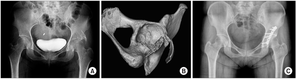
Fig. 1 A 39-year-old female. Preoperative pelvis anteroposterior (AP) radiograph (A) and 3D computed tomography (B) demonstrate a posterior wall fracture. Radiograph obtained 14 months after surgery demonstrate excellent clinical and radiological results.
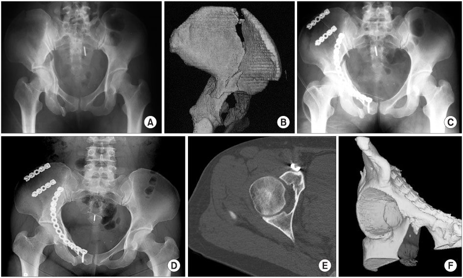
Fig. 2 A38-year-old female. Preoperative pelvic AP radiograph(A)and 3Dcomputed tomography(B)demonstrate bothcolumnfractures. Postoperative pelvis AP radiograph (C) demonstrates an imperfect reduction and secure fixation through the ilioinguinal approach. Radiograph (D) and CT scan (E, F) obtained 39 months after surgery demonstrate good results.
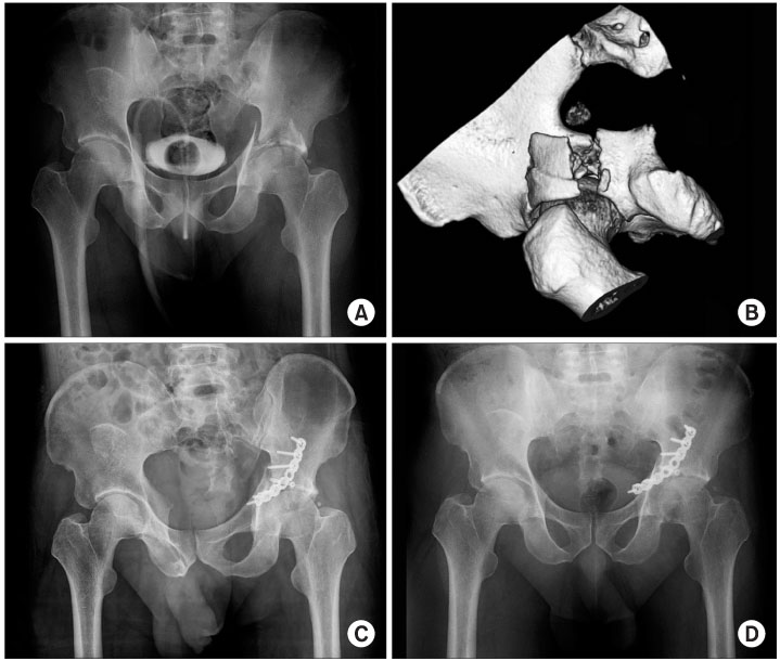
Fig. 3 A 41-year-old male. Preoperative pelvis AP radiograph( A)and 3Dcomputed tomography (B) demonstrates a transeverse fracture. Postoperative pelvis AP radiograph (C) demonstrates poor reduction through the Kocher-Langenbeck approach. Radiograph (D) obtained 17 months postoperatively demonstrate poor results.
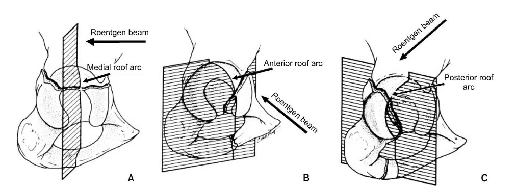
Fig. 4 "Roof arc" measurement, as described by Matta et al. (A) The medial roof arc was measured on anteroposterior view. (B) The anteriorroof arc was measured ona 45-degree angle obturator oblique view. (C)The posteriorroof arc was measured ona 45-degree angle iliac oblique view.
Reference
-
1. Borrelli J Jr, Goldfarb C, Ricci W, Wagner JM, Engsberg JR. Functional outcome after isolated acetabular fractures. J Orthop Trauma. 2002. 16:73–81.
Article2. Browner , Jupiter , Levine , Trafton . Surgical treatment of acetabular fractures. Skeletal trauma. 2003. 2nd ed. Philadelphia: Saunders;1109–1149.3. Carmack DB, Moed BR, McCarroll K, Freccero D. Accuracy of detecting screw penetration of the acetabulum with intraoperative fluoroscopy and computed tomography. J Bone Joint Surg Am. 2001. 83:1370–1375.
Article4. Chiu FY, Chen CM, Lo WH. Surgical treatment of displaced acetabular fractures - 72 cases followed for 10 (6-14) years. Injury. 2000. 31:181–185.
Article5. Choi KS, Chung ES, Kim MK, Son YI. Surgical treatment of the posterior wall fracture of acetabulum with posterior hip dislocation. J Korean Fracture Soc. 1996. 9:525–532.6. d'Aubigne RM, Postel M. Functional results of hip arthroplasty with acrylic prosthesis. J Bone Joint Surg Am. 1954. 36:451–475.7. Judet R, Judet J, Letournel E. Fractures of the acetabulum: classification and surgical approaches for open reduction. Preliminary report. J Bone Joint Surg Am. 1964. 46:1615–1646.8. Min BW, Kang CS, Song KS, Kang CH, Park JW. Cable fixation method for displaced acetabular fracture. J Korean Fracture Soc. 1996. 9:574–582.9. Kim CK, Ahn BW, Lee SG, et al. Surgical and conservative treatment of acetabular fractures. J Korean Fracture Soc. 2003. 16:309–318.
Article10. Kregor PJ, Templeman D. Associated injuries complicating the management of acetabular fractures: review and case studies. Orthop Clin North Am. 2002. 33:73–95.11. Letournel E. The treatment of acetabular fractures through the ilioinguinal approach. Clin Orthop Relat Res. 1993. 292:62–67.
Article12. Letournel E, Judet R. Fractures of the acetabulum. 1993. 2nd ed. Berlin: Springer-Verlag;363–397.13. Letournel E. Diagnosis and treatment of nonunions and malunions of acetabular fractures. Orthop Clin North Am. 1990. 21:769–788.
Article14. Liebergall M, Mosheiff R, Low J, Goldvirt M, Matan Y, Segal D. Acetabular fracture. Clinical outcome of surgical treatment. Clin Orthop Relat Res. 1999. 366:205–216.15. Marsh JL, Buckwalter J, Gelberman R, et al. Articular fracture: does an anatomic reduction really change the result? J Bone Joint Surg Am. 2002. 84:1259–1271.16. Matta JM. Fractures of the acetabulum: accuracy of reduction and clinical results in patients managed operatively within three weeks after the injury. J Bone Joint Surg Am. 1996. 78:1632–1645.17. Matta JM, Anderson LM, Epstein HC, Hendricks P. Fractures of the acetabulum. A retrospective analysis. Clin Orthop Relat Res. 1986. 205:230–240.18. Matta JM, Merritt PO. Displaced acetabular fractures. Clin Orthop Relat Res. 1988. 230:83–97.
Article19. Mears DC, Velyvis JH, Chang CP. Displaced acetabular fractures managed operatively : indicators of outcome. Clin Orthop Relat Res. 2003. 407:173–186.20. Michael RB, Tornetta P III. Orthopaedic knowledge update. 2005. 3rd ed. Orthopaedic Trauma Association;259–280.21. Moed BR, Carr SE, Gruson KI, Watson JT, Craig JG. Computed tomographic assessment of fractures of the posterior wall of the acetabulum after operative treatment. J Bone Joint Surg Am. 2003. 85:512–522.
Article22. Moed BR, WillsonCarr SE, Watson JT. Results of operative treatment of fractures of the posterior wall of the acetabulum. J Bone Joint Surg Am. 2002. 84:752–758.
Article23. Montgomery KD, Potter HG, Helfet DL. Magnetic resonance venography to evaluate the deep venous system of the pelvis in patients who have an acetabular fracture. J Bone Joint Jurg Am. 1995. 77:1639–1649.
Article24. Norris BL, Hahn DH, Bosse MJ, Kellam JF, Sims SH. Intraoperative fluoroscopy to evaluate fracture reduction and hardware placement during acetabular surgery. J Orthop Trauma. 1999. 13:414–417.
Article25. Pantazopoulos T, Mousafiris C. Surgical treatment of central acetabular fractures. Clin Orthop Relat Res. 1989. 246:57–64.
Article26. Pennal GF, Davidson J, Garside H, Plewes J. Result of treatment of acetabular fractures. Clin Orthop Relat Res. 1980. 151:115–123.27. Rice J, Kaliszer M, Dolan M, Cox M, Khan H, McElwain JP. Comparison between clinical and radiologic outcome measures after reconstruction of acetabular fractures. J Orthop Trauma. 2002. 16:82–86.
Article28. de Ridder VA, de Lange S, Kingma L, Hogervorst M. Results of 75 consecutive patients with an acetabular fracture. Clin Orthop Relat Res. 1994. 305:53–57.
Article29. Rowe CR, Lowell JD. Prognosis of fracture of the acetabulum. J Bone Joint Surg Am. 1961. 43:30–59.30. Russell GV Jr, Nork SE, Chip Routt ML Jr. Perioperative complications associated with operative treatment of acetabular fractures. J Trauma. 2001. 51:1098–1103.
Article31. Starr AJ, Watson JT, Reinert CM, et al. Complication s following the "T extensile" approach: a modified extensile approach for acetabular fracture surgery-report of forty-three patients. J Orthop Trauma. 2002. 16:535–542.32. Stockle U, Hoffmann R, Sudkamp NP, Reindl R, Haas NP. Treatment of complex acetabular fractures through a modified extended iliofemoral approach. J Orthop Trauma. 2002. 16:220–230.33. Stover MD, Morgan SJ, Bosse MJ, et al. Prospective comparison of contrast-enhanced computed tomography versus magnetic resonance venography in the detection of occult deep vein thrombosis in patients with pelvic and acetabular fractures. J Orthop Trauma. 2002. 16:613–621.34. Vrahas MS, Widding KK, Thomas KA. The effects of simulated transverse, anterior column, and posterior column fractures of the acetabulum on the stability of the hip joint. J Bone Joint Surg Am. 1999. 81:966–974.
Article
- Full Text Links
-
- Actions
-
Cited
- CITED
-
- Close
- Share
-
- Similar articles
-
- Analysis of Predictors of Results after Surgical Treatment of Acetabular Fractures
- Surgical Treatment of Displaced Acetabular Fractures - focused on Complications after open reduction -
- Surgical Treatment of Displaced Acetabular Fractures
- Surgical Treatment of Acetabular Posterior Wall Fracture with Hip Arthroscopy: A Case Report
- Anterior Approach for the Acetabular Fractures
