Korean J Ophthalmol.
2009 Dec;23(4):306-308. 10.3341/kjo.2009.23.4.306.
Seborrheic Keratosis of the Conjunctiva: A Case Report
- Affiliations
-
- 1Department of Ophthalmology, The Institute of Vision Research, Yonsei University College of Medicine, Seoul, Korea. eungkkim@yuhs.ac
- 2Department of Pathology, Yonsei University College of Medicine, Seoul, Korea.
- KMID: 754769
- DOI: http://doi.org/10.3341/kjo.2009.23.4.306
Abstract
- Seborrheic keratosis is a benign epithelial neoplasia that occurs mainly in the skin of the eyelids and face. We describe a case of seborrheic keratosis of the conjunctiva confirmed by histopathology. A 72-year-old man presented with a recurrent conjunctival mass involving the nasal side of his right eye. Clinically, a diagnosis of conjunctival papilloma was made, and a mass excision was performed. The histopathological analysis evidenced a conjunctival-covering epithelium with papillomatous changes and irregular acanthosis, at the expense of a proliferation of basaloid cells. In addition, the lesion exhibited multiple pseudohorn cysts containing keratin. With the above findings, a diagnosis of conjunctival seborrheic keratosis was established. The occurrence of seborrheic keratosis on the conjunctiva is rare. In this case, seborrheic keratosis was confirmed by pathologic report despite its similar appearance with papilloma. Seborrheic keratosis should be considered in the differential diagnosis of conjunctival lesions.
Keyword
MeSH Terms
Figure
-
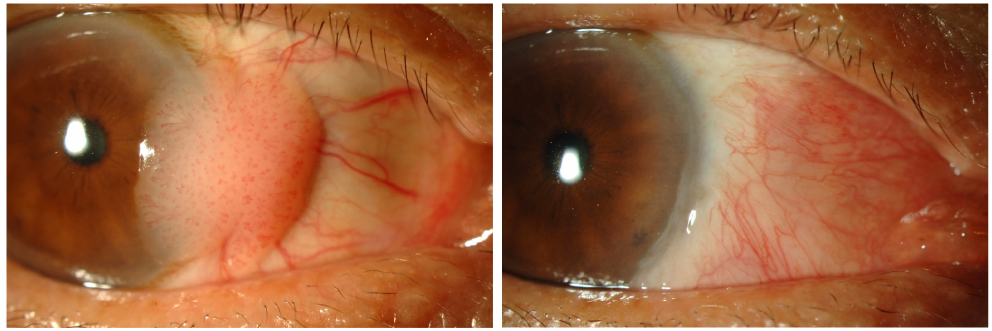
Fig. 1 (A) Photograph showing a juxta-limbal mass over the nasal conjunctiva and invading the cornea. (B) Photograph showing post-operative findings; no mass recurrence was noted.
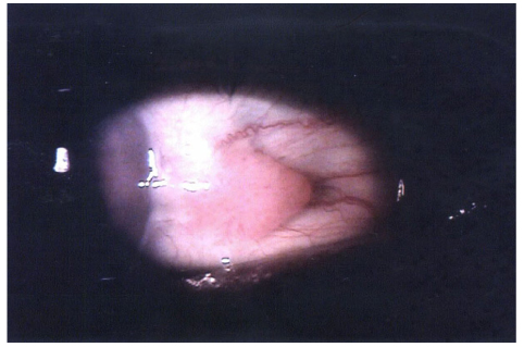
Fig. 2 Photograph from another hospital showing a juxta-limbal mass over the nasal conjunctiva prior to mass excision.

Fig. 3 (A) A histopathologic specimen of the tumor shows acanthosis of uniform basaloid cells with an overlying conjunctival epithelium (H&E, ×40). (B) Pseudohorn cysts containing keratin are seen in the epithelium (H&E, ×100). These histologic features are consistent with seborrheic keratosis.
Reference
-
1. Duke-Elder S. System of Ophthalmology. 1974. Vol. XIII, part I. London: Kimpton;406.2. Albert DM, Jackobiec FA. Principles and Practice of Ophthalmology: Clinical Practice. 1994. Vol. 3. Philadelphia: Saunders;1716–1717.3. Moschella SL, Hurley HJ. Dermatology. 1992. Vol. 2:3rd ed. Philadelphia: Saunders;1722–1723.4. Yanoff M, Fine BS. Ocular Pathology: A Text and Atlas. 1982. 2nd ed. Philadelphia: Harper & Row;241–245.5. Champion RH, Burton JL, Ebling FJ, editors. Rook/Wilkinson/Ebling Textbook of Dermatology. 1992. Vol. 2:5th ed. Oxford: Blackwell Scientific;1465–1467.6. Tseng SH, Chen YT, Huang FC, Jin YT. Seborrheic keratosis of conjunctiva simulating a malignant melanoma: an immunocytochemical study with impression cytology. Ophthalmology. 1999. 106:1516–1520.7. Jain AK, Sukhiha J, Radotra B, Malhota V. Seborrheic keratosis of the conjunctiva. Indian Journal of Ophthalmology. 2004. 52:154–155.8. Gulias-Canizo R, Aranada-Rabago J, Rodriguez-Reyes AA. Seborrheic keratosis of conjunctiva: a case report. Arch Soc Esp Oftalmol. 2006. 81:217–220.9. Seregard S. Conjunctival melanoma. Surv Ophthalmol. 1998. 42:321–350.10. Paridaens ADA, Minassian DC, McCartney ACE, Hungerford JL. Prognostic factors in primary malignant melanoma of the conjunctiva: a clinicopathological study of 256 cases. Br J Ophthalmol. 1994. 78:252–259.11. Yanoff M, Fine BS. Ocular Pathology: A Text and Atlas. 1982. 2nd ed. Philadelphia: Harper & Row;800–804.12. Tsambaos D, Monastirli A, Kapranos N, et al. Detection of human papillomavirus DNA in nongenital seborrheic keratoses. Arch Dermatol Res. 1995. 287:612–615.13. Cascajo CD, Reichel M, Sanchez JL. Malignant neoplasia associated with seborrheic keratoses: an analysis of 54 cases. Am J Dermatopathol. 1996. 18:278–282.14. LeBoit PE, Burg G, Weedon D, Sarasain A, editors. World Health Organization Classification of Tumours: Pathology and Genetics of Skin Tumours. 2006. Lyon: IARC Press;41–43.
- Full Text Links
-
- Actions
-
Cited
- CITED
-
- Close
- Share
-
- Similar articles
-
- A Case of Basal Cell Carcinoma Arising in Seborrheic Keratosis
- A Case of Bowen's Disease Arising in Seborrheic Keratosis
- A Case of Seborrheic Keratosis Distributed as an Agminated form
- Bowen Disease Arising in Recurred Seborrheic Keratosis after Incomplete Removal
- Secondary Localized Cutaneous Amyloidosis in Seborrheic Keratosis
