A Case of Recurrent Schneiderian Papilloma of the Lacrimal Sac Invading the Nasal Cavity
- Affiliations
-
- 1Department of Ophthalmology, Dongsan Medical Center, Keimyung University, Daegu, Korea. changsd@dsmc.or.kr
- 2Department of Pathology, Dongsan Medical Center, Keimyung University, Daegu, Korea.
- KMID: 754725
- DOI: http://doi.org/10.3341/kjo.2009.23.2.100
Abstract
- A 44-year-old man presented with a history of chronic epiphora, discharge from the right eye, and a palpable mass in the medial canthal area. Irrigation of the lacrimal system revealed bloody discharge. Orbital magnetic resonance imaging (MRI) showed a well-defined heterogeneous enhanced mass filling the lacrimal sac and upper nasolacrimal duct (NLD). A wide excision and surgical biopsy were performed. Histopathology showed the tumor to be an exophytic Schneiderian papilloma with moderate to severe dysplasia. Three months later, the mass was found to be invading the nasal cavity through the NLD. Endoscopic histopathological evaluation confirmed that it was identical to the originally identified papilloma.
MeSH Terms
-
Adult
Biopsy
Diagnosis, Differential
Endoscopy
Eye Neoplasms/*pathology/surgery
Follow-Up Studies
Humans
Lacrimal Apparatus/*pathology
Magnetic Resonance Imaging
Male
Nasal Mucosa/*pathology
Neoplasm Invasiveness/*pathology
Neoplasm Recurrence, Local/*pathology/surgery
Nose Neoplasms/*pathology/surgery
Papilloma/*pathology/surgery
Figure
-
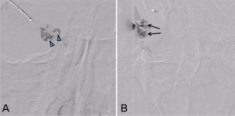
Fig. 1 Dacryocystography shows an uneven, mottled density of contrast media, evidence of a soft tissue mass (A: blue arrow, B: black arrow) at the right lacrimal sac.

Fig. 2 Orbital MRI before surgery. (A) A well-defined mass lesion (arrow) at the right lacrimal sac (T2WI, sagittal view, fat saturation enhancement). (B) A heterogeneous enhancementin the lesion that extends anteriorly adjacent to the right facial soft tissues without evidence of bony structure destruction (T1WI, axial view, Gadolinium enhancement).

Fig. 3 Orbital MRI, 3 months after surgery. (A) A small enhancing recurred lesion (thick arrow) is seen in the right nasolacrimal sac (T1WI, axial view, Gadolinium enhancement). (B) A recurred mass (thin arrow) invading the nasal cavity via the inferior meatus (T1WI, coronal view, Gadolinium enhancement).
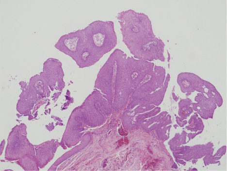
Fig. 4 Histological examination reveals a fungiform mass with projecting finger-like proliferation of the epithelium. The tumor has an exophytic growth pattern with branching fibrovascular stalks covered by an epithelial layer (Hematoxylin-eosin stain, ×40).
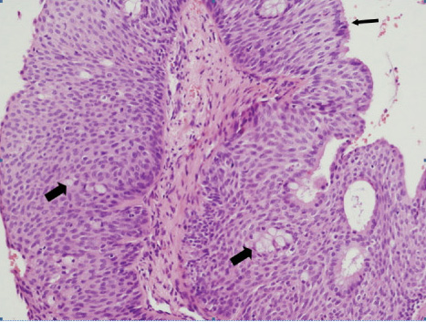
Fig. 5 Transitional-type epithelial lining containing goblet cells (thick arrow). Occasionally, a single layer of columnar ciliated respiratory epithelial cells (thin arrow) is seen to line the surface of the hyperplastic transitional cells (Hematoxylin-eosin stain, ×200).
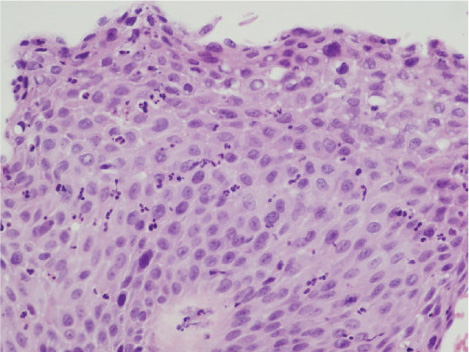
Fig. 6 A partial severe dysplasia in the papilloma (Hematoxylineosin stain ×600).
Reference
-
1. Batsakis JG, Suarez P. Schneiderian papillomas and carcinomas: A review. Adv Anat Pathol. 2001. 8:53–64.2. Batsakis JG. Nasal (Schneiderian) papillomas. Ann Otol Rhinol Laryngol. 1981. 90:190–191.3. Ryan SJ, Font RL. Primary epithelial neoplasms of the lacrimal sac. Am J Ophthalmol. 1973. 76:73–88.4. Ridolfi RL, Lieberman PH, Erlandson RA, Moore OS. Schneiderian papillomas: a clinicopathologic study of 30 cases. Am J Surg Pathol. 1977. 1:43–53.5. Choi KH, Yoon BJ. A case of squamous cell papilloma of the lower canaliculus. J Korean Ophthalmol Soc. 1982. 23:179–181.6. Lee JB, Huh K, Lee TS. A case of squamous cell papilloma of the lacrimal sac. J Korean Ophthalmol Soc. 1991. 32:89–94.7. Valenzuela AA, McNab AA, Selva D, et al. Clinical features and management of tumor affecting the lacrimal drainage apparatus. Ophthal Plast Reconstr Surg. 2006. 22:96–101.8. Flanagan JC, Stokes DP. Lacrimal sac tumors. Ophthalmology. 1978. 85:1282–1287.9. Veirs ER. The lacrimal system First International Symposium. 1971. St. Louis: C.V. Mosby Co;81.10. Parmar DN, Rose GE. Management of lacrimal sac tumours. Eye. 2003. 17:599–606.11. Ashton N, Choyce DP, Fison LG. Carcinoma of the lacrimal sac. Br J Ophthalmol. 1951. 35:366–376.12. Hyams VJ. Papillomas of the nasal cavity and paranasal sinuses. A clinicopathological study of 315 cases. Ann Otol Rhinol Laryngol. 1971. 80:192–206.13. Shan CL, Woo JK, vanHasselt CA. Endoscopic resection of inverted papilloma of nose and paranasal sinuses. J Laryngol Otol. 1998. 112:758–764.
- Full Text Links
-
- Actions
-
Cited
- CITED
-
- Close
- Share
-
- Similar articles
-
- Microorganisms in Conjunctival Sac, Lacrimal Sac and Nasal Cavity
- A Case of An Inverted Papilloma Originating From a Lacrimal Sac
- Schneiderian Papillomas A Clinicopathologic Study of 27 Cases
- Schneiderian Papillomas A Clinicopathologic Study of 27 Cases
- A Case of Oncocytic Schneiderian Papilloma in the Maxillary Sinus
