Evaluation of a New Scoring System for Retinal Nerve Fiber Layer Photography Using HRA1 in 964 Eyes
- Affiliations
-
- 1Institute of Vision Research, Department of Ophthalmology, Yonsei University College of Medicine, Seoul, Korea. youngjhong@gmail.com
- 2Department of Ophthalmology, Soonchunhyang University College of Medicine, Seoul, Korea.
- KMID: 754639
- DOI: http://doi.org/10.3341/kjo.2007.21.4.216
Abstract
- PURPOSE: To evaluate retinal nerve fiber layer (RNFL) defect by a new scoring system for RNFL photography using the Heidelberg Retina Angiograph 1 (HRA1). METHODS: This retrospective study included 128 healthy eyes and 836 primary open-angle glaucoma eyes. The RNFL photography using HRA1 was interpreted using a new scoring system, and correlated with visual field indices of standard automated perimetry (SAP). Using the presence of RNFL defect, darkness, width, and location, we established the new scoring system of RNFL photos. RESULTS: The mean RNFL defect score I in the early, moderate, severe, and control groups were 7.3, 9.2, 10.4, and 3.6, respectively. The mean RNFL defect score II in the early, moderate, severe, and control groups were 14.5, 28.5, 43.4, and 3.4, respectively. Correlations between the RNFL defect score II and the mean deviation of SAP was the strongest of the various combinations (r=-0.675, P<.001). CONCLUSIONS: Using a new scoring system, we propose a method for semi-quantitative interpretation of RNFL photographs. This scoring system may be helpful to distinguish between normal and glaucomatous eyes, and the score is associated with the severity of visual field loss.
MeSH Terms
-
Equipment Design
Female
Fluorescein Angiography/*instrumentation
Fundus Oculi
Glaucoma, Open-Angle/*classification/pathology/physiopathology
Humans
Male
Middle Aged
Nerve Fibers/*pathology
Perimetry
Photography/*instrumentation
Reproducibility of Results
Research Design/*statistics & numerical data
Retinal Ganglion Cells/*pathology
Retrospective Studies
Severity of Illness Index
Visual Fields
Figure
-
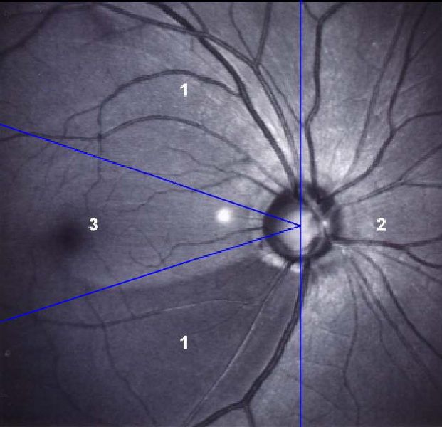
Fig. 1 Location scores of retinal nerve fiber layer defect scoring system. Score "1" represents inferotemporal or superotemporal area, "2" represents nasal area, and "3" represents temporal area.

Fig. 2 (a) Correlations between the retinal nerve fiber layer defect score I and mean deviation of standard automated perimetry(SAP). Y=6.192-0.251 X, r=-0.528, P<0.001. (b) Correlations between the retinal nerve fiber layer defect score II and mean deviation of SAP. Y=9.515-1.772 X, r=-0.675, P<0.001. MD=mean deviation; RNFL=retinal nerve fiber layer.
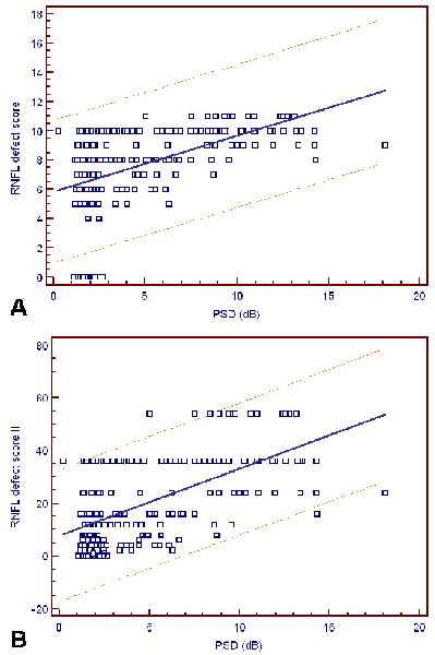
Fig. 3 (a) Correlations between the retinal nerve fiber layer defect score I and pattern standard deviation of standard automated perimetry (SAP). Y=5.841+0.380 X, r=0.481, P<0.001. (b) Correlations between the retinal nerve fiber layer (RNFL) defect score II and pattern standard deviation (PSD) of standard automated perimetry (SAP). Y=7.642+2.542 X, r=0.581, P<0.001. PSD= pattern standard deviation; RNFL=retinal nerve fiber layer.

Fig. 4 Correlations between the retinal nerve fiber layer defect score II and mean deviation of standard automated perimetry in early stage glaucoma and control group. Y=2.849-2.386 X, r=-0.313, P<0.001. MD=mean deviation; RNFL=retinal nerve fiber layer.
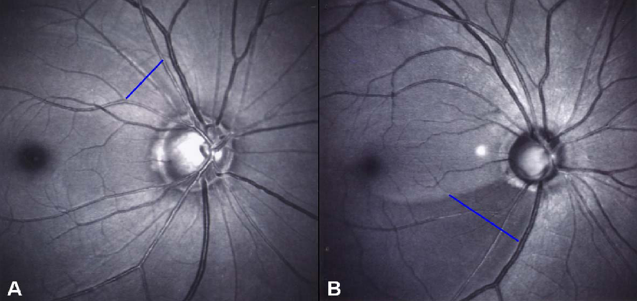
Fig. 5 Examples of interpretation of the retinal nerve fiber layer photographs using HRA1. (a) Early stage glaucoma patient (MD -1.59 dB, PSD 2.60 dB). RNFL defect, darkness, width, and location score are 1 (suspicious defect), 1 (slight), 1 (< 45(), and 1 (superotemporal), respectively. RNFL defect score I is 4, and RNFL defect score II is 1. (b) Moderate advanced glaucoma patient (MD -6.77 dB, PSD 10.81 dB). RNFL defect, darkness, width, and location score are 2 (definite defect), 2 (moderate), 2 (45° - 90° ), and 1 (inferotemporal), respectively. RNFL defect score I is 7, and RNFL defect score II is 8. Blue lines indicated RNFL defect area.
Cited by 1 articles
-
Comparison of Blue and Green Confocal Scanning Laser Ophthalmoscope Imaging to Detect Retinal Nerve Fiber Layer Defects
Joo Young Joung, Won June Lee, Byung Ro Lee
Korean J Ophthalmol. 2019;33(2):131-137. doi: 10.3341/kjo.2018.0075.
Reference
-
1. Hoyt WF, Frisen L, Newman NM. Fundoscopy of nerve fiber layer defects in glaucoma. Invest Ophthalmol. 1973. 12:814–829.2. Quigley HA, Katz J, Derick RJ, et al. An evaluation of optic disc and nerve fiber layer examinations in monitoring progression of early glaucoma damage. Ophthalmology. 1992. 99:19–28.3. Sommer A. Retinal nerve fiber layer. Am J Ophthalmol. 1995. 120:665–667.4. Sommer A, Katz J, Quigley HA, et al. Clinically detectable nerve fiber atrophy precedes the onset of glaucomatous field loss. Arch Ophthalmol. 1991. 109:77–83.5. Sommer A, Miller NR, Pollack I, et al. The nerve fiber layer in the diagnosis of glaucoma. Arch Ophthalmol. 1977. 95:2149–2156.6. Niessen AG, van den Berg TJ, Langerhorst CT, Bossuyt PM. Grading of retinal nerve fiber layer with a photographic reference set. Am J Ophthalmol. 1995. 120:577–586.7. Niessen AG, van den Berg TJ. Evaluation of a reference set based grading system for retinal nerve fiber layer photographs in 1941 eyes. Acta Ophthalmol Scand. 1998. 76:278–282.8. Sommer A, Quigley HA, Robin AL, et al. Evaluation of nerve fiber layer assessment. Arch Ophthalmol. 1984. 102:1766–1771.9. Hong S, Ahn H, Ha SJ, Yeom HY, Seong GJ, Hong YJ. Early Glaucoma Detection Using the Humphrey Matrix Perimeter, GDx VCC, Stratus OCT, and Retinal Nerve Fiber Layer Photography. Ophthalmology. 2007. 114:210–215.10. Anderson DR. Automated static perimetry. 1992. 1st ed. St Louis: Mosby;123.11. Hodapp E, Parrish RK II, Anderson DR. Clinical decisions in glaucoma. 1993. 1st ed. St. Louis: Mosby;1–204.12. Caprioli J, Miller JM, Sears M. Quantitative evaluation of the optic nerve head in patients with unilateral visual field loss from primary open-angle glaucoma. Ophthalmology. 1987. 94:1484–1487.13. Garway-Heath DF, Hitchings RA. Quantitative evaluation of the optic nerve head in early glaucoma. Br J Ophthalmol. 1998. 82:352–361.14. Jonas JB, Gusek GC, Naumann GO. Optic disc morphometry in chronic primary open-angle glaucoma. I. Morphometric intrapapillary characteristics. Graefes Arch Clin Exp Ophthalmol. 1988. 226:522–530.15. Pederson JE, Anderson DR. The mode of progressive disc cupping in ocular hypertension and glaucoma. Arch Ophthalmol. 1980. 98:490–495.
- Full Text Links
-
- Actions
-
Cited
- CITED
-
- Close
- Share
-
- Similar articles
-
- Reproducibility of Retinal Nerve Fiber Layer Thickness Evaluation by Nerve Fiber Analyzer
- Early Detection of Glaucoma with Retinal Nerve Fiber Layer Photograph
- Biometry of Retinal Nerve Fiber Layer Thickness by NFA
- Influence of Diabetes Mellitus on the Retinal Ne rve Fiber Layer Thickness Measurement by Nerve Fiber Analyzer
- A Case of Retinal Herniation through Peripapillary Pit Resulting in Retinal Nerve Fiber Layer Defect
