Arteriovenous Sheathotomy for Persistent Macular Edema in Branch Retinal Vein Occlusion
- Affiliations
-
- 1Hangil Eye Hospital, Incheon, Korea.
- 2Kangbuk Samsung Hospital, Department of Ophthalmology, Sungkyunkwan University College of Medicine, Seoul, Korea. ssjeye @yahoo.co.kr
- KMID: 754584
- DOI: http://doi.org/10.3341/kjo.2006.20.4.210
Abstract
- PURPOSE: To evaluate the efficacy of arteriovenous (AV) sheathotomy with internal limiting membrane peeling for persistent or recurrent macular edema after intravitreal triamcinolone injection and/or laser photocoagulation in branch retinal vein occlusion. METHODS: Twenty-two eyes with branch retinal vein occlusion (BRVO) with recurrent macular edema underwent vitrectomy with AV sheathotomy and internal limiting membrane peeling. All eyes had previous intravitreal triamcinolone injection and/or laser photocoagulation for macular edema. The best corrected visual acuity (BCVA), fluorescein angiography and optical coherence tomography (OCT) before and after surgery were compared. RESULTS: The mean preoperative BCVA (log MAR) were 0.79+/-0.29 and postoperative BCVA (log MAR) at 3 months was 0.57+/-0.33. And improvement of visual acuity > or =2 lines was observed in 10 eyes (45%). The mean preoperative fovea thickness measured by OCT was 595.22+/-76.83 micrometer (510-737 micrometer) and postoperative fovea thickness was 217.60+/-47.33 micrometer (164-285 micrometer). CONCLUSIONS: Vitrectomy with AV sheathotomy can be one treatment option for the patients with recurrent macular edema in BRVO.
MeSH Terms
-
Treatment Outcome
Tomography, Optical Coherence
Retrospective Studies
Retinal Vein Occlusion/*complications/diagnosis/surgery
Ophthalmologic Surgical Procedures/*methods
Middle Aged
Male
Macular Edema, Cystoid/diagnosis/etiology/*surgery
Macula Lutea/*surgery
Humans
Fundus Oculi
Follow-Up Studies
Fluorescein Angiography
Female
Figure
-
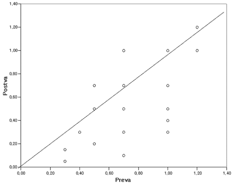
Fig. 1 The relationship between the preoperative visual acuity and postoperative visual acuity. The improvement of visual acuity is statistically significant (p<0.01, Chi squared test). Preva: preoperative visual acuity (log MAR), Postva: postoperative visual acuity (log MAR)

Fig. 2 Preoperative and postoperative fundus photography and OCT in a 64 year-old patient with BRVO. (visual acuity 0.1) (A) Widespread retina hemorrhage and exudation. (B) Preoperative fluorescein angiography of nonperfusion area after 3 months after laser and IVTA treatment. (C) Color fundus photography 1 year after surgery. (visual acuity 0.3). (D) Preoperative OCT showed cystoid macular edema with central foveal thickness 552 µm. E. Postoperative OCT with decreased macular edema with central foveal thickness 214 µm.
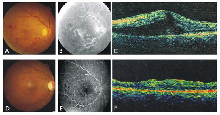
Fig. 3 Preoperative and postoperative fundus photography and fluorescein angiography of a 67 year-old patient with inferotemporal BRVO (visual acuity 0.2). (A) Preoperative fluorescein angiography showing retina ischemia in inferotemporal area including macula 3 months after IVTA. (B) Postoperative fluorescein angiography at 1 year after surgery (visual acuity 0.4) with a lack of blocked fluorescence and minimal leakage. (C) Preoperative OCT with central fovea thickness 637 µm. (D) Postoperative OCT 15 months after surgery with fovea thickness 183 µm.
Cited by 1 articles
-
Macular Ischemia Correlated with Final Visual Outcome in Retinal Vein Occlusion Patients
Gwang Myung Noh, Ji Eun Lee, Ki Yup Nam, Seung Uk Lee, Sang Joon Lee
J Korean Ophthalmol Soc. 2014;55(10):1493-1498. doi: 10.3341/jkos.2014.55.10.1493.
Reference
-
1. Orth DH, Patz A. Retinal branch vein occlusion. Surv Ophthalmol. 1978. 22:357–376.2. Branch Vein Occlusion Study Group. Argon laser scatter photocoagulation for prevention of neovascularization and vitreous hemorrhage in branch vein occlusion. Arch Ophthalmol. 1986. 104:31–41.3. Jonas JB, Akkoyun I, Kamppeter B, Kreissig I, Degenring RF. Branch retinal vein occlusion treated by intravitreal triamcinolone acetonide. Eye. 2005. 19:65–71.4. Yepremyan M, Wertz FD, Tivnan T, et al. Early treatment of cystoid macular edema secondary to branch retinal vein occlusion with intravitreal triamcinolone acetonide. Ophthalmic Surg Lasers Imaging. 2005. 36:30–36.5. Mester U, Dillinger P. Vitrectomy with arteriovenous decompression and internal limiting membrane dissection in branch retinal vein occlusion. Retina. 2002. 22:740–746.6. Le Rouic JF, Bejjani RA, Rumen F, et al. Adventitial sheathotomy for decompression of recent onset branch retinal vein occlusion. Graefes Arch Clin Exp Ophthalmol. 2001. 239:747–751.7. Opremcak EM, Bruce RA. Surgical decompression of branch retinal vein occlusion via arteriovenous crossing sheathotomy: a prospective review of 15 cases. Retina. 1999. 19:1–5.8. Han DP, Bennett SR, Williams DF, Dev S. Arteriovenous crossing dissection without separation of the retina vessels for treatment of branch retinal vein occlusion. Retina. 2003. 23:145–151.9. Fujii GY, de Juan E Jr, Humayun MS. Improvements after sheathotomy for branch retinal vein occlusion documented by optical coherence tomography and scanning laser ophthalmoscope. Ophthalmic Surg Lasers Imaging. 2003. 34:49–52.10. Mason J 3rd, Feist R, White M Jr, et al. Sheathotomy to decompress branch retinal vein occlusion: a matched control study. Ophthalmology. 2004. 111:540–545.11. Charbonnel J, Glacet-Bernard A, Korobelnik JF, et al. Management of branch retinal vein occlusion with vitrectomy and arteriovenous adventitial sheathotomy, the possible role of surgical posterior vitreous detachment. Graefes Arch Clin Exp Ophthalmol. 2004. 242:223–228.12. Zhao J, Sastry SM, Sperduto RD, et al. Arteriovenous crossing patterns in branch retinal vein occlusion: The Eye Disease Case-control Study Group. Ophthalmology. 1993. 100:423–428.13. Branch Vein Occlusion Study Group. Argon laser scatter photocoagulation for macular edema in branch vein occlusion. Arch Ophthalmol. 1984. 98:271–282.14. Weinberg D, Dodwell DG, Fern SA. Anatomy of arteriovenous crossings in branch retinal vein occlusion. Am J Ophthalmol. 1990. 109:298–302.15. Ozkris A, Evereklioglu C, Erkilic K, Illhan O. The efficacy of intravitreal triamcinolone acetonide on macular edema in branch retinal vein occlusion. Eur J Ophthalmol. 2005. 15:96–101.16. Stefansson E, Novack RL, Hatchell DL. Vitrectomy prevents retinal hypoxia in branch retinal vein occlusion. Invest Ophthalmol Vis Sci. 1990. 31:284–289.17. Osterloh MD, Charles S. Surgical decompression of branch retinal vein occlusion. Arch Ophthalmol. 1988. 106:1469–1471.18. Yamamoto S, Saito W, Yagi F, et al. Vitrectomy with or without arteriovenous adventitial sheathotomy for macular edema associated with branch retinal vein occlusion. Am J Ophthalmol. 2004. 138:907–914.19. Lakhanpal RR, Javaheri M, Ruiz-Garcia H, et al. Transvitreal limited arteriovenous-crossing manipulation without vitrectomy for complicated branch retinal vein occlusion using 25-gauge instrumentation. Retina. 2005. 25:272–280.20. Garcia-Arumi J, Martinez-Castillo V, Boixadera A, et al. Management of macular edema in branch retinal vein occlusion with sheathotomy and recombinant tissue plasminogen activator. Retina. 2004. 24:530–540.21. Mandelcorn MS, Nrusimhadevara RK. Internal limiting membrane peeling for decompression of macular edema in retinal vein occlusion: a report of 14 cases. Retina. 2004. 24:348–355.
- Full Text Links
-
- Actions
-
Cited
- CITED
-
- Close
- Share
-
- Similar articles
-
- Five Cases of Arteriovenous Adventitial Sheathotomy for Branch Retinal Vein Occlusion
- Arteriovenous Adventitial Sheathotomy for the Treatment of Macular Edema Associated with Branch Retinal Vein Occlusion
- The Effect of Vitrectomy and Arteriovenous Adventitial Sheathotomy for Branch Retinal Vein Occlusion
- Long-term Visual Outcome of Arteriovenous Adventitial Sheathotomy on Branch Retinal Vein Occlusion Induced Macular Edema
- Retinal Arteriolar Changes in a Patient with Branch Retinal Vein Occlusion
