A Case of Enterococcus Faecalis Endophthalmitis with Corneal Ulcer
- Affiliations
-
- 1Department of Ophthalmology, Mokdong Hospital, Ewha Womans University College of Medicine, Seoul, Korea.
- KMID: 754384
- DOI: http://doi.org/10.3341/kjo.2004.18.2.175
Abstract
- Although there have been a few reported cases of Enterococcal endophthalmitis, this is an unusual case of endophthalmitis complicated with corneal ulcer caused by Enterococcus faecalis. A 67-year-old male patient with diabetes mellitus underwent secondary intraocular lens implantation. Post-operative recovery was uneventful until a wound rupture was noted 3 weeks after the operation. On day 12 after the repair of the wound, endophthalmitis accompanied by wound necrosis and a fullthickness corneal ulcer was detected. His vision was light perception, and Enterococcus faecalis was identified by culture in samples of conjunctival sac, anterior chamber and vitreous humor. After 3 rounds of intravitreal antibiotics injection, the vitreous opacity disappeared on ultrasonographic finding but corneal opacity and corneal neovascularization still remained.
MeSH Terms
-
Aged
Anti-Bacterial Agents/administration & dosage
Corneal Ulcer/diagnosis/drug therapy/*microbiology
Endophthalmitis/diagnosis/drug therapy/*microbiology
Enterococcus faecalis/drug effects/*isolation & purification
*Eye Infections, Bacterial/diagnosis/drug therapy/microbiology
Gram-Positive Bacterial Infections/diagnosis/drug therapy/*microbiology
Humans
Lens Implantation, Intraocular
Male
Microbial Sensitivity Tests
Surgical Wound Infection/diagnosis/drug therapy/*microbiology
Treatment Outcome
Figure
-

Fig. 1 (A) Ultrasonographic finding of the right eye shows vitreous opacity on admission day. (B) Ultrasonographic finding shows vitreous opacity is slightly increased in spite of the double intravitreal antibiotics injection (3 days after the second intravitreal injection). (C) Ultrasonographic finding shows vitreous opacity is decreased on final follow up findings (1 month after the first intravitreal injection).
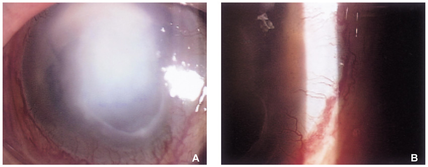
Fig. 2 (A) Anterior segment photograph shows epithelial defect and corneal opacity still remain after the vitreous opacity was subsided. (B) Corneal neovascularization is also noted at the limbus.
Cited by 4 articles
-
A Case of Bilateral Endogenous Enterococcus Faecalis Endophthalmitis in Liver Abscess
Ji Hye Song, In Young Chung, Jong Moon Park
J Korean Ophthalmol Soc. 2007;48(9):1291-1296. doi: 10.3341/jkos.2007.48.9.1291.Prognostic Factors in Patients With Endogenous Endophthalmitis
Jeong Ho Hwang, Nam Chun Cho
J Korean Ophthalmol Soc. 2009;50(6):858-863. doi: 10.3341/jkos.2009.50.6.858.A Case of Bilateral Endogenous Endophthalmitis in a Streptococcus pneumoniae Meningitis Patient
Min Soo Lee, Moo Hwan Chang
J Korean Ophthalmol Soc. 2013;54(2):370-374. doi: 10.3341/jkos.2013.54.2.370.Clinical and Microbiological Analysis of Gram-Positive Bacterial Keratitis, a 15-Year Review
Mi Rae Kim, Sang Bumm Lee
J Korean Ophthalmol Soc. 2014;55(10):1432-1444. doi: 10.3341/jkos.2014.55.10.1432.
Reference
-
1. Mundy LM, Sham DF, Gilmore MS. Relationship between enterococcal virulence and antimicrobial resistance. Clin Microbiol Rev. 2000. 13:513–522.2. Ejdervik-Lindblad B, Lindberg M, Hakansson E. Enterococcal endophthalmitis following cataract extraction treated with ampicillin intravitreally. Acta Ophthalmol. 1992. 70:842–843.3. Uchio E, Inamura M, Okada K, Hatano H, Saeki K, Ohno S. A case of endogenous Enterococcus faecalis endophthalmitis. Jpn J Ophthalmol. 1992. 36:215–221.4. Fraser SG, Ohri R. Endophthalmitis caused by Enterococcus faecalis. Eye. 1995. 9:535–536.5. Bae JH, Lee SS. A case of Enterococcus faecalis endophthalmitis following ECCE. J Korean Ophthalmol Soc. 1994. 35:66–70.6. Kim US, Yu SY, Kwak HW. Two cases of Enterococcus faecalis endophthalmitis. J Korean Ophthalmol Soc. 2003. 44:523–528.7. Han DP, Wisniewski SR, Wilson LA, Barza M, Vine A, Doft BH, Kelsey SF. The Endophthalmitis Vitrectomy Study Group. Spectrum and susceptibilities of microbiologic isolates in the endophthalmitis vitrectomy study. Am J Ophthalmol. 1996. 122:1–17.8. Endophthalmitis Vitrectomy Study Group. Microbiologic factors and visual outcome in the endophthalmitis vitrectomy study. Am J Ophthalmol. 1996. 122:830–846.9. Driebe WT, Mandelbaum S, Foster RK, Schwartz LK, Culbertson WW. Pseudophakic endophthalmitis, diagnosis and management. Ophthalmology. 1986. 93:442–448.10. Callegan MC, Booth MC, Jett BD, Gilmore MS. Pathogenesis of gram-positive bacterial endophthalmitis. Infect Immun. 1999. 67:3348–3356.11. Phillips WB, Tasman WS. Postoperative endophthalmitis in association with diabetes mellitus. Ophthalmology. 1994. 101:508–518.12. Jett BD, Huycke MM, Gilmore MS. Virulence of enterococci. Clin Microbiol Rev. 1994. 7:462–478.13. Callegan MC, Engelbert M, Parke DW II, Jett BD, Gilmore MS. Bacterial endophthalmitis: epidemiology, therapeutics, and bacterium-host interactions. Clin Microbiol Rev. 2002. 15:111–124.14. Jett BD, Atkuri RV, Gilmore MS. Enterococcus faecalis localization in experimental endophthalmitis : role of plasmid-encoded aggregation substance. Infect Immun. 1998. 66:843–848.15. Jett BD, Jenson HG, Atkuri RV, Gilmore MS. Evaluation of therapeutic measures for treating endophthalmitis caused by isogenic toxin-producing and toxin-nonproducing Enterococcus faecalis strains. Invest Ophthalmol Vis Sci. 1992. 33:1650–1656.16. Endophthalmitis Vitrectomy Study Group. Results of the endophthalmitis vitrectomy study: a randomized trial of immediate vitrectomy and of intravenous antibiotics for the treatment of postoperative bacterial endophthalmitis. Arch Ophthalmol. 1995. 113:1479–1496.17. Doft BH, Misniewski SR, Kelsey SF, Fitzgerald SG. The Endophthalmitis Vitrectomy Study Group. Diabetes and postoperative endophthalmitis in the endophthalmitis vitrectomy study. Arch Ophthalmol. 2001. 119:650–656.18. Cameron JA, Badr IA, Miguel Risco J, Abboud E, Gonnah el-S. Endophthalmitis cluster from contaminated donor corneas following penetrating keratoplasty. Can J Ophthalmol. 1998. 33:8–13.
- Full Text Links
-
- Actions
-
Cited
- CITED
-
- Close
- Share
-
- Similar articles
-
- A Case of Bilateral Endogenous Enterococcus Faecalis Endophthalmitis in Liver Abscess
- Recurrent Enterococcus faecalis Endophthalmitis
- A Case of Corneal Ulcer by Alcaligenes Faecalis
- Analysis of Genetic Mutations in Quinolone Resistance and Virulence Factor Gene Profile of Enterococcus faecalis
- A Case of Enterococcus Faecalis Endophthalmitis after Phacoemulsification
