MR Imaging of Shaken Baby Syndrome Manifested as Chronic Subdural Hematoma
- Affiliations
-
- 1Department of Radiology, Hallym University Sacred Heart Hospital, Kyungki-do, Korea. leeyul@www.hallym.or.kr
- KMID: 754103
- DOI: http://doi.org/10.3348/kjr.2001.2.3.171
Abstract
- Shaken baby syndrome (SBS) is a form of child abuse that can cause significant head injuries, of which subdural hematoma (SDH) is the most common manifestation. We report the MRI findings of chronic SDH in three cases of SBS, involving two-, three- and eight-month-old babies. The SDH signal was mostly low on T1-weighted images and high on T2-weighted images, suggesting chronic SDH. In chronic SDH, a focal high signal on T1-weighted images was also noted, suggesting rebleeding. Contrast-enhanced MRI revealed diffuse dural enhancement.
MeSH Terms
Figure
-
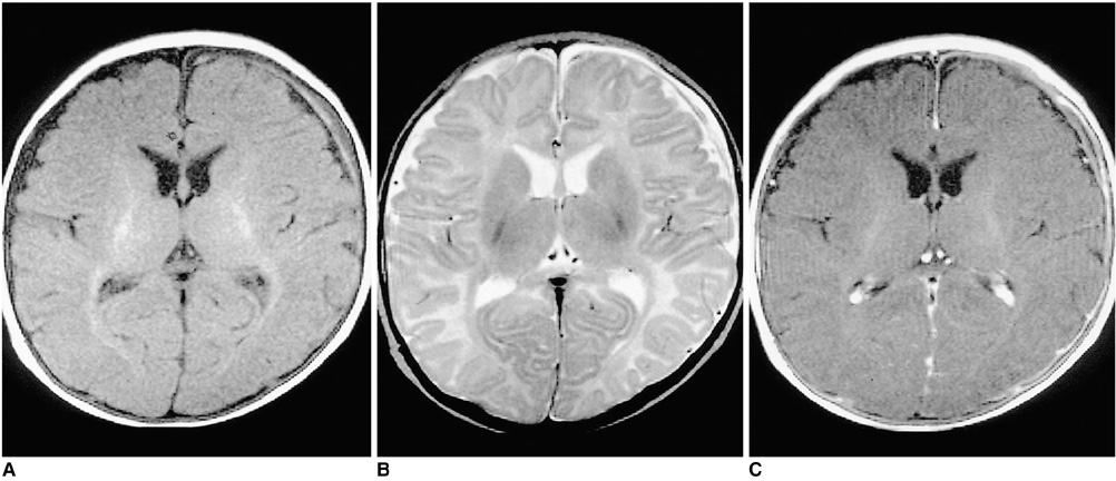
Fig. 1 Chronic subdural hematoma (SDH) in a three-month-old female patient. A. T1-weighted image shows mainly low-signal SDH, with a high signal focus in the left frontal area. B. On a T2-weighted image the signal intensity of the chronic SDH is mainly high, with a focal area of low intensity. C. Contrast-enhanced T1-weighted image shows overlying linear dural enhancement.
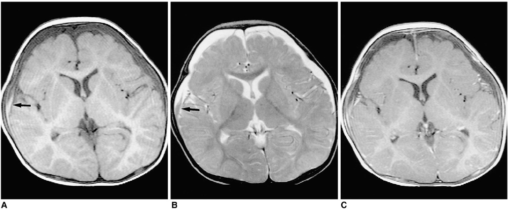
Fig. 2 Chronic SDH in an eight-month-old male patient. A. T1-weighted image shows low-signal SDH in both frontal areas. A high signal area, suggesting subacute hemorrhage, may also be observed in the right frontal area (arrow). B. On a T2-weighed image, the signal intensity of the SDH is mainly high, though there is a focal area of low intensity (arrow). C. Contrast-enhanced T1-weighted image shows diffuse linear dural enhancement.
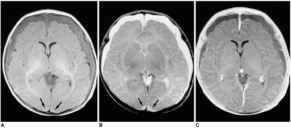
Fig. 3 Chronic SDH in a two-month-old female patient. A. T1-weighted image shows low (though higher than CSF)-signal SDH in both cerebral hemispheres and the posterior fossa. In addition, a high signal area, suggesting subacute hemorrhage, can be seen in the occipital areas (arrows). B. T2-weighted image reveals high signal intensity SDHs in both frontoparietal and occipital areas (arrows). C. Contrast-enhanced T1-weighted image demonstrates diffuse linear dural enhancement.
Reference
-
1. Cox LA. The shaken baby syndrome: diagnosis using CT and MRI. Radiol Technol. 1996. 67:513–520.2. Petitti N, Williams DW III. CT and MR imaging of nonaccidental pediatric head trauma. Acad Radiol. 1998. 5:215–223.3. Sato Y, Yuh WTC, Smith WL, Alexander RC, Kao SCS, Ellerbroek CJ. Head injury in child abuse: evaluation with MR imaging. Radiology. 1989. 173:653–657.4. Duheime AC, Gennarelli TA, Tiubault LE, Bruce DA, Margulies SS, Wiser R. The shaken baby syndrome: A clinical, pathological and biochemical study. J Neurosurg. 1987. 66:409–415.5. Hearley MN, Sonntag VKH, Rekate HL, Murphy A. The infant whiplash-shake injury syndrome: a clinical and pathologic study. Neurosurgery. 1989. 24:536–540.6. Couser S. Shaken baby syndrome. J Pediatr Health Care. 1993. 7:238–239.7. Brown GR, Runyan DK. Diagnosing child maltreatment. N C Med J. 1994. 55:404–408.8. Kleinman PK. Diagnostic imaging in infant abuse. AJR. 1990. 155:703–712.9. Osborn AG. Diagnostic neuroradiology. 1994. St. Louis: Mosby;205–212.10. Budenz DL, Farber MG, Mirchandani HG, Park H, Rorke LB. Ocular and optic nerve hemorrhages in abused infants with intracranial injuries. Ophthalmology. 1994. 101:559–565.
