Brain MRI Findings of Carbon Disulfide Poisoning
- Affiliations
-
- 1Department of Radiology, Wonjin Green Hospital, Kangwon-do, Korea. sskim@kangwon.ac.kr
- 2Department of Radiology, Kangwon National University College of Medicine, Kangwon-do, Korea.
- 3Department of Environmental Health, School of Public Health, Seoul National University, Seoul, Korea.
- 4Department of Occupational Medicine, Wonjin Green Hospital, Kangwon-do, Korea.
- 5Department of Family Medicine, Wonjin Green Hospital, Kangwon-do, Korea.
- KMID: 754057
- DOI: http://doi.org/10.3348/kjr.2002.3.3.158
Abstract
OBJECTIVE
To evaluate the findings of brain MRI in patients with carbon disulfide poisoning.
MATERIALS AND METHODS
Ninety-one patients who had suffered carbon disulfide poisoning [male:female=87:4; age, 32-74 (mean 53.3) years] were included in this study. To determine the extent of white matter hyperintensity (Grade 0-V) and lacunar infarction, T2-weighted MR imaging of the brain was performed.
RESULTS
T2-weighted images depicted white matter hyperintensity in 70 patients (76.9%) and lacunar infarcts in 27 (29.7%).
CONCLUSION
In these patients, the prevalent findings at T2-weighted MR imaging of the brain were white matter hyperintensity and lacunar infarcts. Disturbance of the cardiovascular system by carbon disulfide might account for these results.
Keyword
MeSH Terms
Figure
-
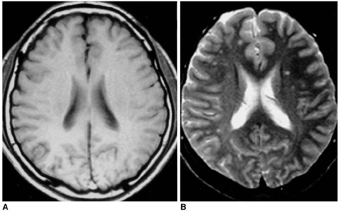
Fig. 1 A 34-year-old man exposed to carbon disulfide for nine years, but with no accompanying disease. T1- (A) and T2-weighted (B) axial images depict between 10 and 20 grade-II focal white matter lesions in the fontal lobe.
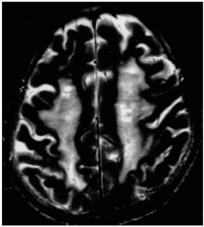
Fig. 2 A 64-year-old man with hypertension after exposure to carbon disulfide for 13 years. T2-weighted axial image shows a grade-V, diffuse, confluent white matter lesion in the centrum semiovale.
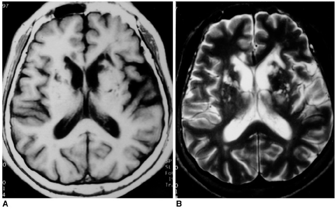
Fig. 3 A 58-year-old man exposed to carbon disulfide for 15 years, but with no accompanying disease. T1- (A) and T2-weighted (B) images indicate that multifocal lacunar infarcts are present in the basal ganglia and genu of the corpus callosum.
Cited by 1 articles
-
Occupational Neurologic Disorders in Korea
Yangho Kim, Kyoung Sook Jeong, Yong-Hun Yun, Myoung-Soon Oh
J Clin Neurol. 2010;6(2):64-72. doi: 10.3988/jcn.2010.6.2.64.
Reference
-
1. Davidson M, Feinleib M. Carbon disulfide poisoning: A review. Am Heart J. 1972. 83:100–114.2. Lee KB, Byoun HJ, Choi TS, Kim SS, Cho WY, Kim HK. Clinical manifestation of chronic carbon disulfide intoxication. Korean J Int Med. 1990. 39:245–251.3. Choi JW, Jang SH. A review of the carbon disulfide poisoning experience in Korea. Korean J Occup Med. 1991. 3:11–20.4. Yang KS, Choi HR, Kim JJ, et al. Study of carbon disulfide intoxication. 1999. Seoul: Korean Ministry of Labor Press.5. Aaserud O, Gierstad L, Nakstad P, et al. Neurological examination, computerized tomography, cerebral blood flow and neurophysiological examination in workers with long-term exposure to carbon disulfide. Toxicology. 1988. 49:277–282.6. Aaserud O, Hommeren OJ, Tvedt B, et al. Carbon disulfide exposure and neurotoxic sequelae among viscose rayon workers. Am J Ind Med. 1990. 18:25–37.7. Sugimura K, Kabashima K, Tatetsu S. Computerized tomography in chronic carbon disulfide poisoning. No to Shinkei. 1979. 31:1245–1253.8. Huang CC, Chu CC, Chen RS, et al. Chronic carbon disulfide encephalopathy. Eur Neurol. 1996. 36:364–368.9. Peters HA, Levine RL, Matthews CGM, et al. Extrapyramidal and other neurologic manifestations associated with carbon disulfide fumigant exposure. Arch Neurol. 1988. 45:537–540.10. Horikoshi T, Yagi S, Fukamachi A. Incidental high-intensity foci in white matter on T2-weighted magnetic resonance imaging. Neuroradiology. 1993. 35:151–155.11. Wojtczak-Jaroszowa J, Kubow S. Carbon monoxide, carbon disulfide, lead and cadmium-four examples of occupational toxic agents linked to cardiovascular disease. Med Hypotheses. 1989. 30:141–150.12. Awad IA, Spetzler RF, Hodak JA, Awad CA, Carey R. Incidental subcortical lesions identified on magnetic resonance imaging in the elderly: I. Correlation with age and cerebrovascular risk factors. Stroke. 1986. 17:1084–1089.13. Fazekas F. Magnetic resonance signal abnormalities in asymptomatic individuals: their incidental and functional correlates. Eur Neurol. 1989. 29:164–168.14. Hendrie HC, Farlow MR, Austrom MG, Edward MK, Williams MA. Foci of increased T2 signal intensity on brain MR scans of healthy elderly subjects. AJNR. 1989. 10:703–707.15. Yetkin FZ, Fischer ME, Papke RA, Haughton VM. Focal hyperintensities in cerebral white matter on MR images of asymptomatic volunteers: correlation with social and medical histories. AJR. 1993. 161:855–858.16. Gerard G, Weisberg LA. MRI periventricular lesions in adults. Neurology. 1986. 36:998–1001.17. Braffman BH, Zimmerman RA, Trojanowski JQ, Gonatas NK, Hickey WF, Schlaepfer WW. Brain MR: pathologic correlation with gross and histopathology. 2. Hyperintense white-matter foci in the elderly. AJR Am J Roentgenol. 1988. 151:559–566.18. Brant-Zawadzki M, Kucharczk W. Brant-Zawadzki M, Norman D, editors. Vascular disease: ischemia. Magnetic resonance imaging of the central nervous system. 1987. New York: Raven;221–234.19. Lee E, Kim MH. Cerebral vasoreactivity by transcranial Doppler in carbon disulfide poisoning cases in Korea. J Korean Med Sci. 1998. 13:645–651.20. Victor M, Ropper AH. Principles of Neuroradiology. 2001. New York: McGraw-Hill;847–851.21. Fisher CM. Lacunes: small, deep cerebral infarcts. Neurology. 1965. 15:774–784.22. Fisher CM. Lacunar strokes and infarcts: a review. Neurology. 1982. 32:871–896.23. Braffman BH, Zimmerman RA, Trojanowski JQ, Gonatas NK, Hickey WF, Schlaepfer WW. Brian MR: pathologic correlation with gross and histopathology. 1. Lacunar infarction and Virchow-Robin spaces. AJR Am J Roentgenol. 1988. 151:551–558.
- Full Text Links
-
- Actions
-
Cited
- CITED
-
- Close
- Share
-
- Similar articles
-
- A review of the carbon disulfide poisoning experiences in Korean
- Clinical observation on patients with chronic carbon disulfide poisoning
- Health status of workers exposed to carbon disulfide at a viscoserayon factory in Korea
- Clinical Analysis of 8 Cases of Chronic Carbon Disulfide Poisoning in Workers Engaged in the Viscose Rayon Industry
- The study of blood carbon disulfide in rats after oral administration of carbon disulfide
