Sclerosing Stromal Tumor of the Ovary: MR-Pathologic Correlation in Three Cases
- Affiliations
-
- 1Department of Radiology, School of Medicine, Catholic University of Daegu. kjjung@cataegu.ac.kr
- 2Department of Radiology, School of Medicine, Keimyung University.
- 3Department of Radiology, School of Medicine, Donga University.
- KMID: 754025
- DOI: http://doi.org/10.3348/kjr.2003.4.3.194
Abstract
- Sclerosing stromal tumor (SST) of the ovary is a very rare sex cord stromal tumor occurring in a younger age group than other types of stromal tumors and most commonly accompanied by menstrual irregularity. Several unique histologic features including pseudolobulation, sclerosis and prominent vascularity are clearly reflected at ultrasonography and MRI. We report the ultrasonographic and MR features of three cases of histologically confirmed SSTs, and relate them to the pathological findings.
Keyword
Figure
-
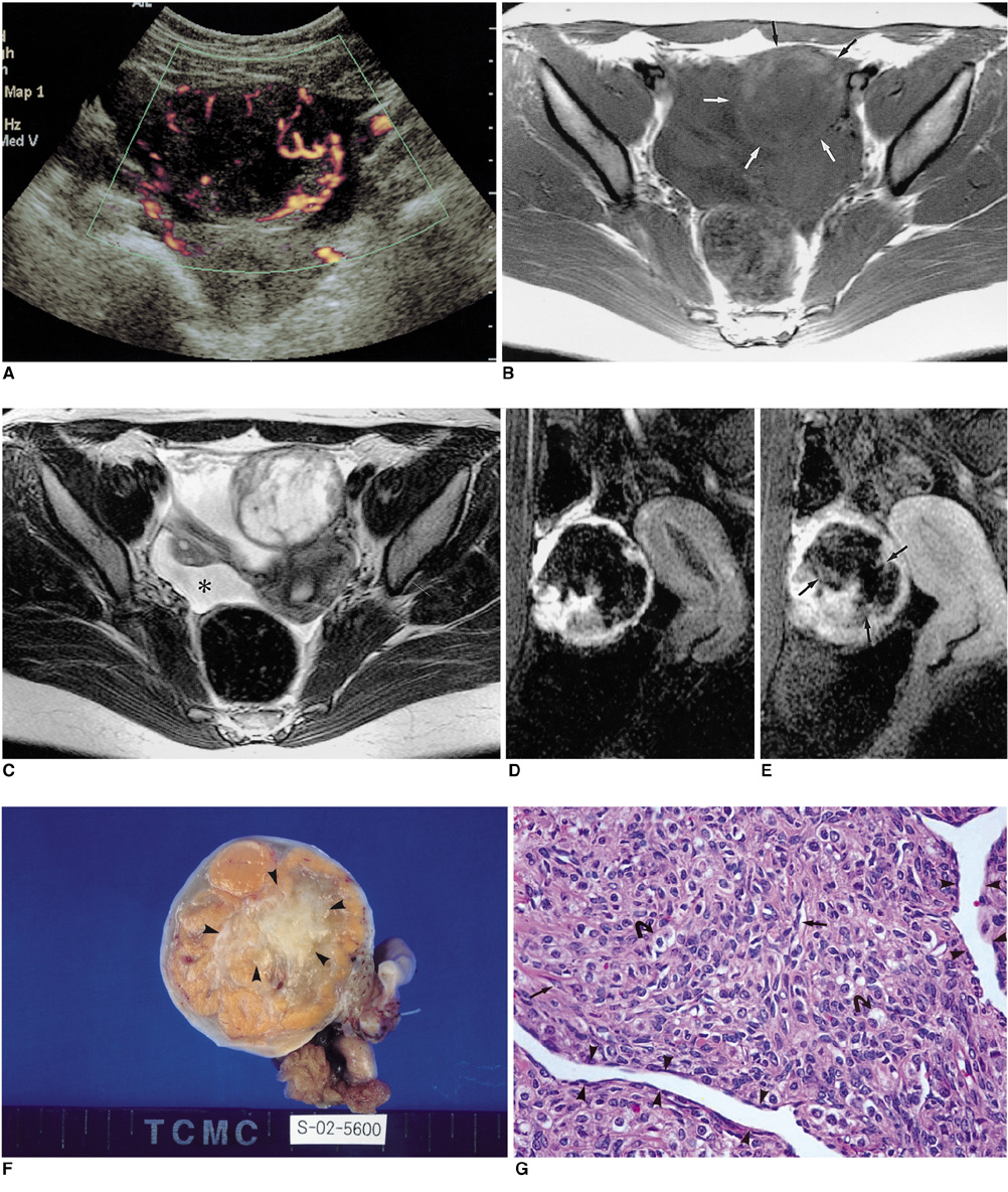
Fig. 1 Sclerosing stromal tumor of the left ovary in a 16-year-old girl. A. Peripheral arc-like vessels with a vertically oriented centripetal vascular network (the so-called "spoke-wheel appearance") in an oval-shaped left adnexal mass are clearly demonstrated at power Doppler ultrasonography. B. Axial T1-weighted MR image depicts a left adnexal mass (arrows). The peripheral portion of the mass is slightly hyperintense relative to muscle, and the central portion is hypointense. C. Axial T2-weighted MR image shows a slightly hyperintense peripheral portion relative to muscle, and a hyperintense central portion with a peripheral low signal rim. Slight ascites is present (*). D. Sagittal gradient-echo (GE) image obtained 40 seconds after the administration of Gd-DTPA reveals strong enhancement of the peripheral portion of the mass. E. Sagittal GE image obtained 140 seconds after demonstrates progressive centripetal enhancement (arrows). F. At the sectioned surface of the tumor, a central whitish edematous and collagenous area (arrowheads) is visible, and this is surrounded by yellowish solid tissue, with a whitish ovarian capsule at its outermost rim. G. Microscopic image of the cellular portion reveals an admixture of fibroblasts (arrows) and rounded vascuolated cells (curved arrows), and prominent thin-walled vessels (arrowheads) are noticeable (H&E staining, ×200).
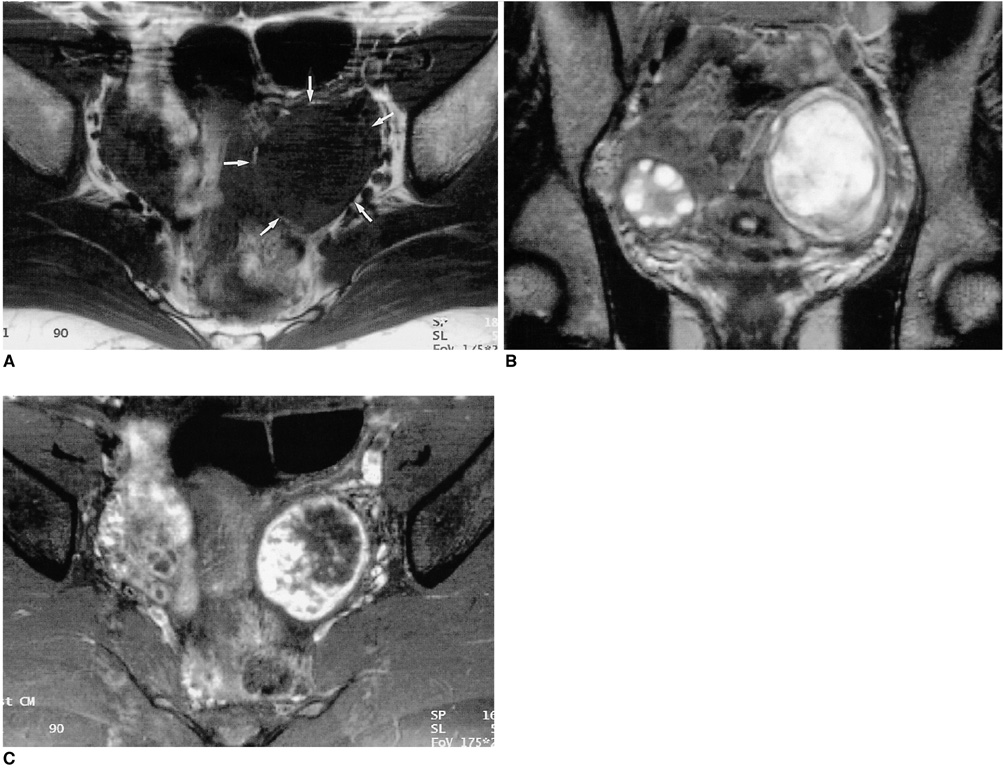
Fig. 2 Sclerosing stromal tumor of the left ovary in a 26-year-old woman. A. Axial T1-weighted MR image demonstrates a round, left adnexal mass (arrows), with a slightly hyperintense peripheral portion relative to muscle and a homogeneous central hypointense area. B. Coronal T2-weighted image reveals a left adnexal mass with a marked hyperintense central portion and slightly hyperintense peripheral portion. The mass is surrounded by a low-signal peripheral rim. C. Gadolinium-enhanced MR image demonstrates strong peripheral nodular enhancement.
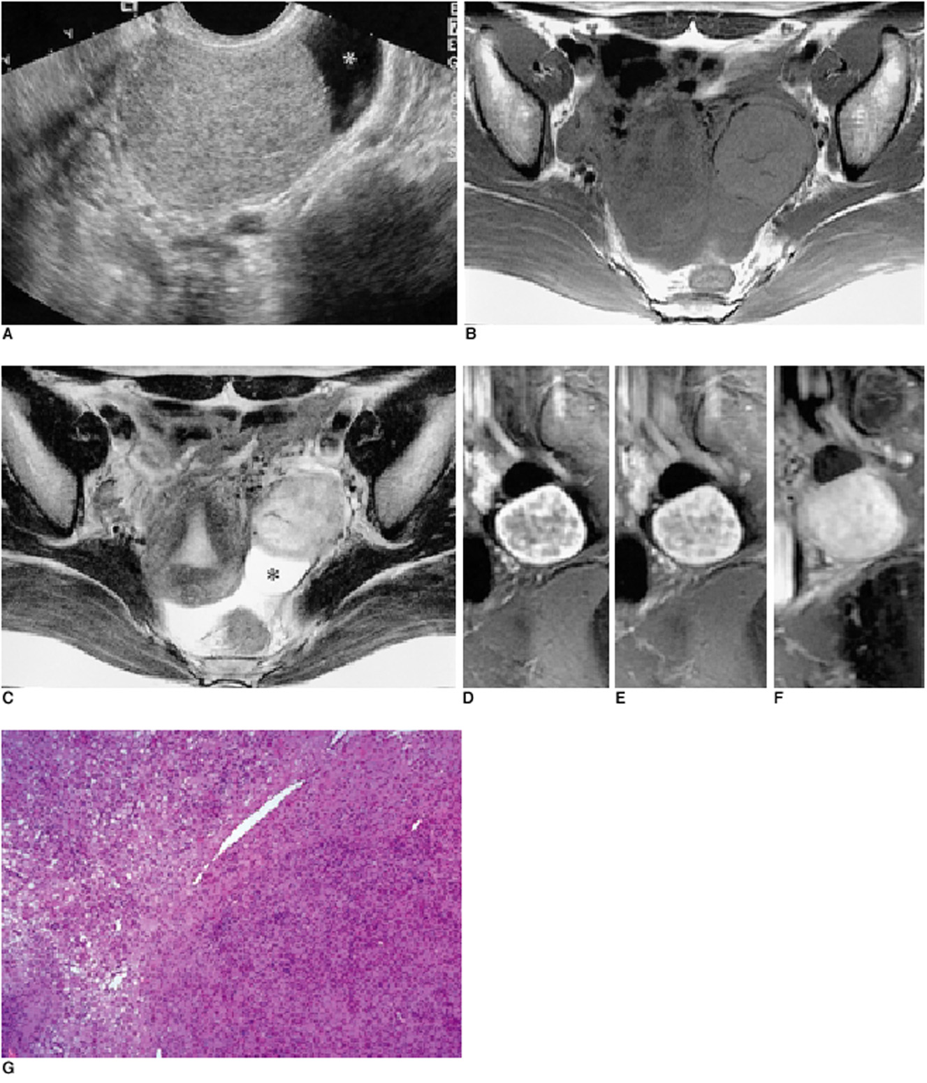
Fig. 3 Sclerosing stromal tumor of the left ovary in a 39-year-old woman. A. Transvaginal ultrasonography reveals a homogeneously echogenic solid mass in the left adnexa. At the left of the mass, a cyst is present (*). B. Axial T1-weighted MR image depicts a left adnexal mass which, relative to muscle, showed slightly high signal intensity. Signal void tubular structures are seen peripherally. C. Axial T2-weighted image of the mass, demonstrating heterogeneous high signal intensity and peripheral signal-void tubular structures. Elongated cysts surround the mass (*), and a cul-de-sac contains a small amount of ascites. D, E, F. Dynamic contrast-enhanced MR images obtained 30 seconds (D), 60 seconds (E), and 5 minutes (F) after the administration of Gd-DTPA reveal, except in small scattered areas of the cyst and cleft, marked, early, peripheral nodular enhancement, and progressive centripetal enhancement. G. Microscopic examination reveals a pseudolobular pattern with cellular nodules separated by less cellular areas of edematous connective tissue (H&E staining, ×100).
Reference
-
1. Ihara N, Togashi K, Todo G, et al. Sclerosing stromal tumor of the ovary: MRI. J Comput Assist Tomogr. 1999. 23:555–557.2. Matsubayashi R, Matsuo Y, Doi J, Kudo S, Matsuguchi K, Sugimori H. Sclerosing stromal tumor of the ovary: radiologic findings. Eur Radiol. 1999. 9:1335–1338.3. Kawauchi S, Tsuji T, Kaku T, Kamura T, Nakano H, Tsuneyoshi M. Sclerosing stromal tumor of the ovary: a clinicopathologic, immunohistochemical, ultrastructural, and cytogenetic analysis with special reference to its vasculature. Am J Surg Pathol. 1998. 22:83–92.4. Lee MS, Cho HC, Lee Y-H, Hong SR. Ovarian sclerosing stromal tumors: gray scale and color Doppler sonographic findings. J Ultrasound Med. 2001. 20:413–417.5. Joja I, Okuno K, Tsunoda M, et al. Sclerosing stromal tumor of the ovary: US, MR, and dynamic MR findings. J Comput Assist Tomogr. 2001. 25:201–206.6. Outwater EK, Wagner BJ, Mannion C, McLarney JK, Kim B. Sex cord-stromal and steroid cell tumors of the ovary. RadioGraphics. 1998. 18:1523–1546.7. Chalvardjian A, Scully RE. Sclerosing stromal tumors of the ovary. Cancer. 1973. 31:664–670.8. Jung SE, Lee JM, Rha SE, Byun JY, Jung JI, Hahn ST. CT and MR imaging of ovarian tumors with emphasis on differential diagnosis. RadioGraphics. 2002. 22:1305–1325.9. Damjanov I, Drobnjak P, Grizelj V, Longhino N. Sclerosing stromal tumor of the ovary: a hormonal and ultrastructural analysis. Obstet Gynecol. 1975. 45:675–679.10. Yuen BH, Robertson I, Clement PB, Mincey EK. Sclerosing stromal tumor of the ovary. Obstet Gynecol. 1982. 60:252–256.
