Acute Crit Care.
2024 Feb;39(1):155-161. 10.4266/acc.2023.01172.
Bedside ultrasonographic evaluation of optic nerve sheath diameter for monitoring of intracranial pressure in traumatic brain injury patients: a cross sectional study in level II trauma care center in India
- Affiliations
-
- 1Department of Anesthesiology and Critical Care, B. J. Government Medical College and Sassoon General Hospital, Pune, India
- KMID: 2555234
- DOI: http://doi.org/10.4266/acc.2023.01172
Abstract
- Background
Optic nerve sheath diameter (ONSD) is an emerging non-invasive, easily accessible, and possibly useful measurement for evaluating changes in intracranial pressure (ICP). The utilization of bedside ultrasonography (USG) to measure ONSD has garnered increased attention due to its portability, real-time capability, and lack of ionizing radiation. The primary aim of the study was to assess whether bedside USG-guided ONSD measurement can reliably predict increased ICP in traumatic brain injury (TBI) patients. Methods: A total of 95 patients admitted to the trauma intensive care unit was included in this cross sectional study. Patient brain computed tomography (CT) scans and Glasgow Coma Scale (GCS) scores were assessed at the time of admission. Bedside USG-guided binocular ONSD was measured and the mean ONSD was noted. Microsoft Excel was used for statistical analysis. Results: Patients with low GCS had higher mean ONSD values (6.4±1.0 mm). A highly significant association was found among the GCS, CT results, and ONSD measurements (P<0.001). Compared to CT scans, the bedside USG ONSD had 86.42% sensitivity and 64.29% specificity for detecting elevated ICP. The positive predictive value of ONSD to identify elevated ICP was 93.33%, and its negative predictive value was 45.00%. ONSD measurement accuracy was 83.16%. Conclusions: Increased ICP can be accurately predicted by bedside USG measurement of ONSD and can be a valuable adjunctive tool in the management of TBI patients.
Figure
-
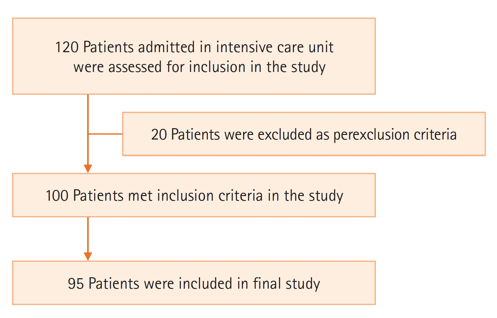
Figure 1. Flowchart of the study.
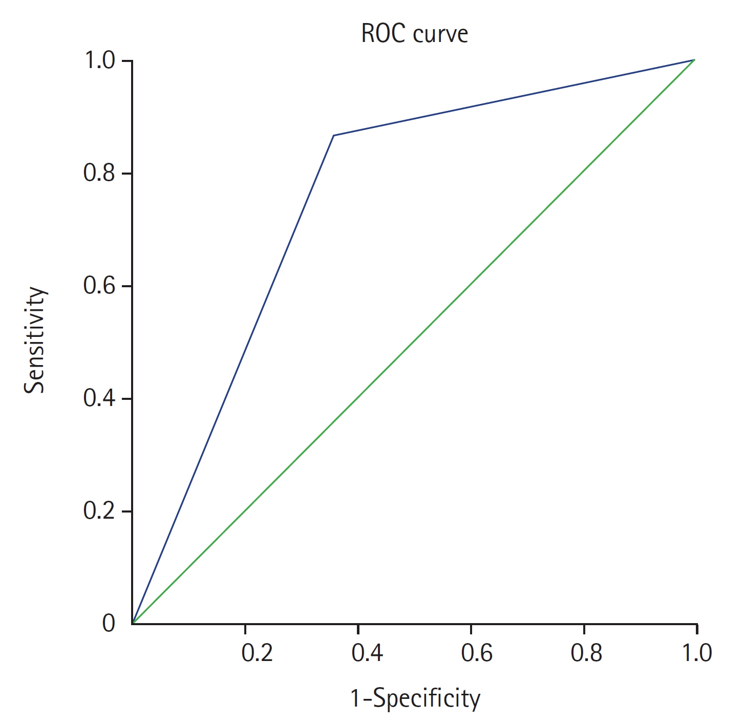
Figure 2. Receiver operating characteristic (ROC) curve analysis of optic nerve sheath diameter.
Reference
-
1. Soldatos T, Karakitsos D, Chatzimichail K, Papathanasiou M, Gouliamos A, Karabinis A. Optic nerve sonography in the diagnostic evaluation of adult brain injury. Crit Care. 2008; 12:R67.
Article2. Moretti R, Pizzi B, Cassini F, Vivaldi N. Reliability of optic nerve ultrasound for the evaluation of patients with spontaneous intracranial hemorrhage. Neurocrit Care. 2009; 11:406–10.
Article3. Moretti R, Pizzi B. Optic nerve ultrasound for detection of intracranial hypertension in intracranial hemorrhage patients: confirmation of previous findings in a different patient population. J Neurosurg Anesthesiol. 2009; 21:16–20.
Article4. Dubourg J, Javouhey E, Geeraerts T, Messerer M, Kassai B. Ultrasonography of optic nerve sheath diameter for detection of raised intracranial pressure: a systematic review and meta-analysis. Intensive Care Med. 2011; 37:1059–68.
Article5. Geeraerts T, Launey Y, Martin L, Pottecher J, Vigué B, Duranteau J, et al. Ultrasonography of the optic nerve sheath may be useful for detecting raised intracranial pressure after severe brain injury. Intensive Care Med. 2007; 33:1704–11.
Article6. Geeraerts T, Merceron S, Benhamou D, Vigué B, Duranteau J. Non-invasive assessment of intracranial pressure using ocular sonography in neurocritical care patients. Intensive Care Med. 2008; 34:2062–7.
Article7. Kimberly HH, Shah S, Marill K, Noble V. Correlation of optic nerve sheath diameter with direct measurement of intracranial pressure. Acad Emerg Med. 2008; 15:201–4.
Article8. Kaur A, Gautam PL, Sharma S, Singh VP, Sharma S. Bedside ultrasonographic assessment of optic nerve sheath diameter as a means of detecting raised intracranial pressure in neuro-trauma patients: a cross-sectional study. Ann Indian Acad Neurol. 2021; 24:63–8.
Article9. Raffiz M, Abdullah JM. Optic nerve sheath diameter measurement: a means of detecting raised ICP in adult traumatic and non-traumatic neurosurgical patients. Am J Emerg Med. 2017; 35:150–3.
Article10. Zhu S, Cheng C, Zhao D, Zhao Y, Liu X, Zhang J. The clinical and prognostic values of optic nerve sheath diameter and optic nerve sheath diameter/eyeball transverse diameter ratio in comatose patients with supratentorial lesions. BMC Neurol. 2021; 21:259.
Article11. Helmke K, Hansen HC. Fundamentals of transorbital sonographic evaluation of optic nerve sheath expansion under intracranial hypertension : II. patient study. Pediatr Radiol. 1996; 26:706–10.
Article12. Yanamandra U, Gupta A, Yanamandra S, Das SK, Patyal S, Nair V. Bedside ultrasonography as an alternative to computed tomography scan for the measurement of optic nerve sheath diameter. J Neurosci Rural Pract. 2018; 9:252–5.
Article13. Tayal VS, Neulander M, Norton HJ, Foster T, Saunders T, Blaivas M. Emergency department sonographic measurement of optic nerve sheath diameter to detect findings of increased intracranial pressure in adult head injury patients. Ann Emerg Med. 2007; 49:508–14.
Article14. Blaivas M, Theodoro D, Sierzenski PR. Elevated intracranial pressure detected by bedside emergency ultrasonography of the optic nerve sheath. Acad Emerg Med. 2003; 10:376–81.
Article15. Gauthey M, Tessaro MO, Breitbart S, Kulkarni AV, Davis AL. Reliability and feasibility of optic nerve point-of-care ultrasound in pediatric patients with ventricular shunts. Childs Nerv Syst. 2022; 38:1289–95.
Article16. Rajajee V, Vanaman M, Fletcher JJ, Jacobs TL. Optic nerve ultrasound for the detection of raised intracranial pressure. Neurocrit Care. 2011; 15:506–15.
Article17. Bittencourt Rynkowski C, Caldas J. Ten good reasons to practice neuroultrasound in critical care setting. Front Neurol. 2022; 12:799421.18. Beckmann U, Gillies DM, Berenholtz SM, Wu AW, Pronovost P. Incidents relating to the intra-hospital transfer of critically ill patients: an analysis of the reports submitted to the Australian Incident Monitoring Study in Intensive Care. Intensive Care Med. 2004; 30:1579–85.
Article19. Aliaga M, Forel JM, De Bourmont S, Jung B, Thomas G, Mahul M, et al. Diagnostic yield and safety of CT scans in ICU. Intensive Care Med. 2015; 41:436–43.
Article20. Caruana M, Culp K. Intrahospital transport of the critically ill adult: a research review and implications. Dimens Crit Care Nurs. 1998; 17:146–56.21. Dinsmore M, Venkatraghavan L. Clinical applications of point-of-care ultrasound in brain injury: a narrative review. Anaesthesia. 2022; 77 Suppl 1:69–77.
Article22. Wang LJ, Chen LM, Chen Y, Bao LY, Zheng NN, Wang YZ, et al. Ultrasonography assessments of optic nerve sheath diameter as a noninvasive and dynamic method of detecting changes in intracranial pressure. JAMA Ophthalmol. 2018; 136:250–6.
Article23. Hirzallah MI, Lochner P, Hafeez MU, Lee AG, Krogias C, Dongarwar D, et al. Quality assessment of optic nerve sheath diameter ultrasonography: scoping literature review and Delphi protocol. J Neuroimaging. 2022; 32:808–24.
Article24. Montorfano L, Yu Q, Bordes SJ, Sivanushanthan S, Rosenthal RJ, Montorfano M. Mean value of B-mode optic nerve sheath diameter as an indicator of increased intracranial pressure: a systematic review and meta-analysis. Ultrasound J. 2021; 13:35.
Article
- Full Text Links
-
- Actions
-
Cited
- CITED
-
- Close
- Share
-
- Similar articles
-
- Comparison of the effects of normal and low blood pressure regulation on the optic nerve sheath diameter in robot assisted laparoscopic radical prostatectomy
- Availability of the Optic Nerve Sheath Diameter Measured by Using Ultrasonography as a Secondary Survey for Patient with Head Injuries in the Emergency Department
- Real-time change of optic nerve sheath diameter after rebleeding of ruptured intracranial dissecting aneurysm
- The Relation between Neurologic Prognosis and Optic Nerve Sheath Diameter Measured in Initial Brain Computed Tomography of Cardiac Arrest and Hanging Patients
- Correlation between Optic Nerve Sheath Diameter Measured by Computed Tomography and Elevated Intracranial Pressure in Patients with Traumatic Brain Injury
