Medial Ankle Impingement Syndrome due to Talar Osteochondroma and Gout Attack: A Case Report
- Affiliations
-
- 1Department of Orthopedic Surgery, Seoul National University Hospital, Seoul, Korea
- 2Department of Orthopedic Surgery, Seoul National University College of Medicine, Seoul, Korea
- KMID: 2553807
- DOI: http://doi.org/10.14193/jkfas.2024.28.1.31
Abstract
- Osteochondromas are benign bone tumors typically found in the metaphyseal region of long bones. These tumors are often asymptomatic and detected incidentally. However, their occurrence in atypical sites such as the talus can pose significant diagnostic and treatment challenges. This report describes a rare case of osteochondroma of the medial tubercle of the talus, which is an unprecedented location based on a review of relevant literature. A 28-year-old male presented with worsening medial ankle pain and limping. Imaging revealed a lesion consistent with osteochondroma contributing to medial ankle impingement syndrome. Uniquely, this case also featured a coinciding gout attack in the ankle joint. Surgical removal of the lesion resulted in significant symptom relief and functional improvement. This case underscores the need to consider rare diagnoses, such as talar osteochondroma, when presented with persistent medial ankle pain and highlights the potential presence of concurrent conditions, such as gout.
Keyword
Figure
-
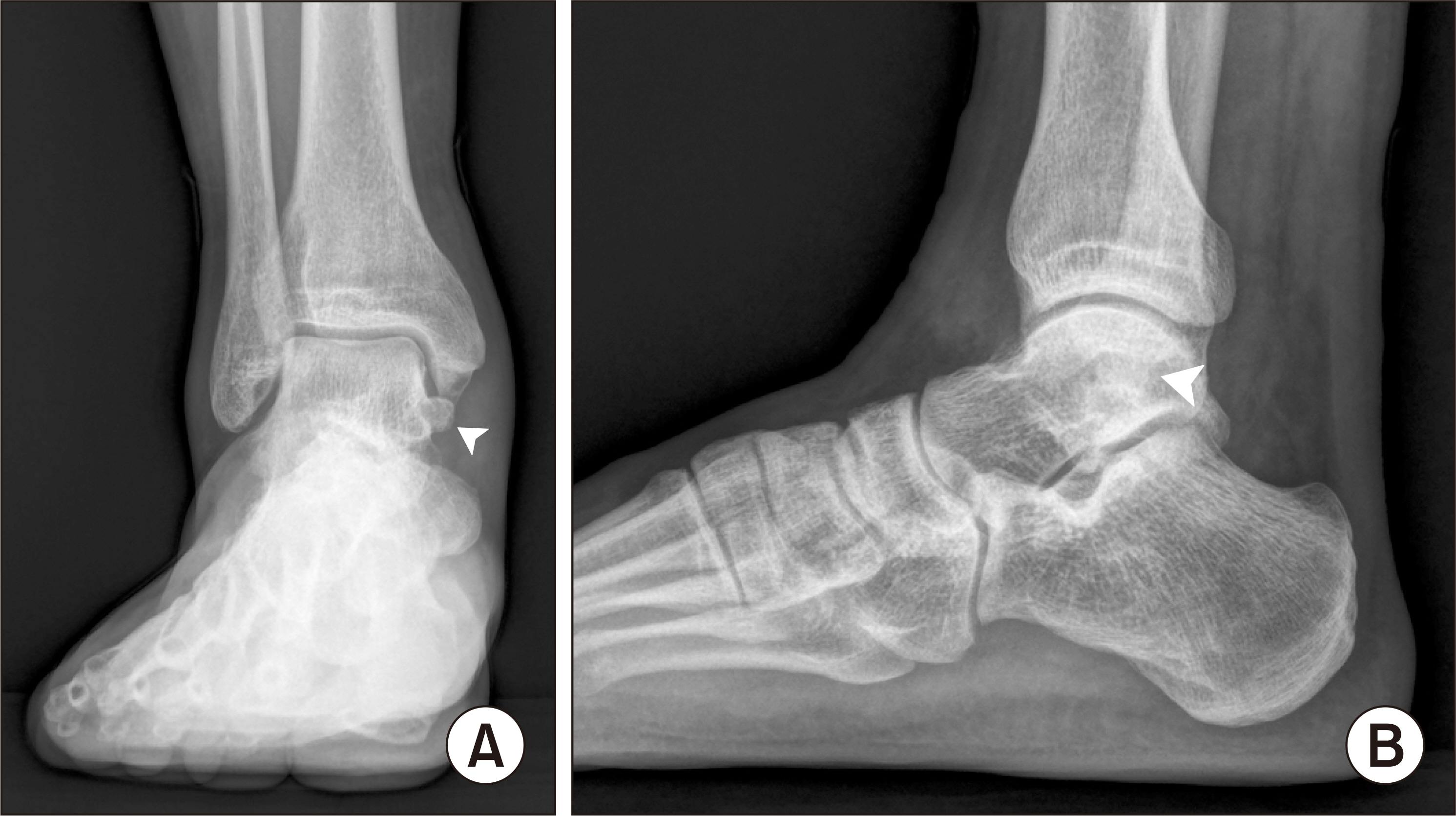
Figure 1 Initial standing ankle (A) anteroposterior and (B) lateral radiographs demonstrating the presence of a bony protrusion (arrowhead) at the medial side of the talus.
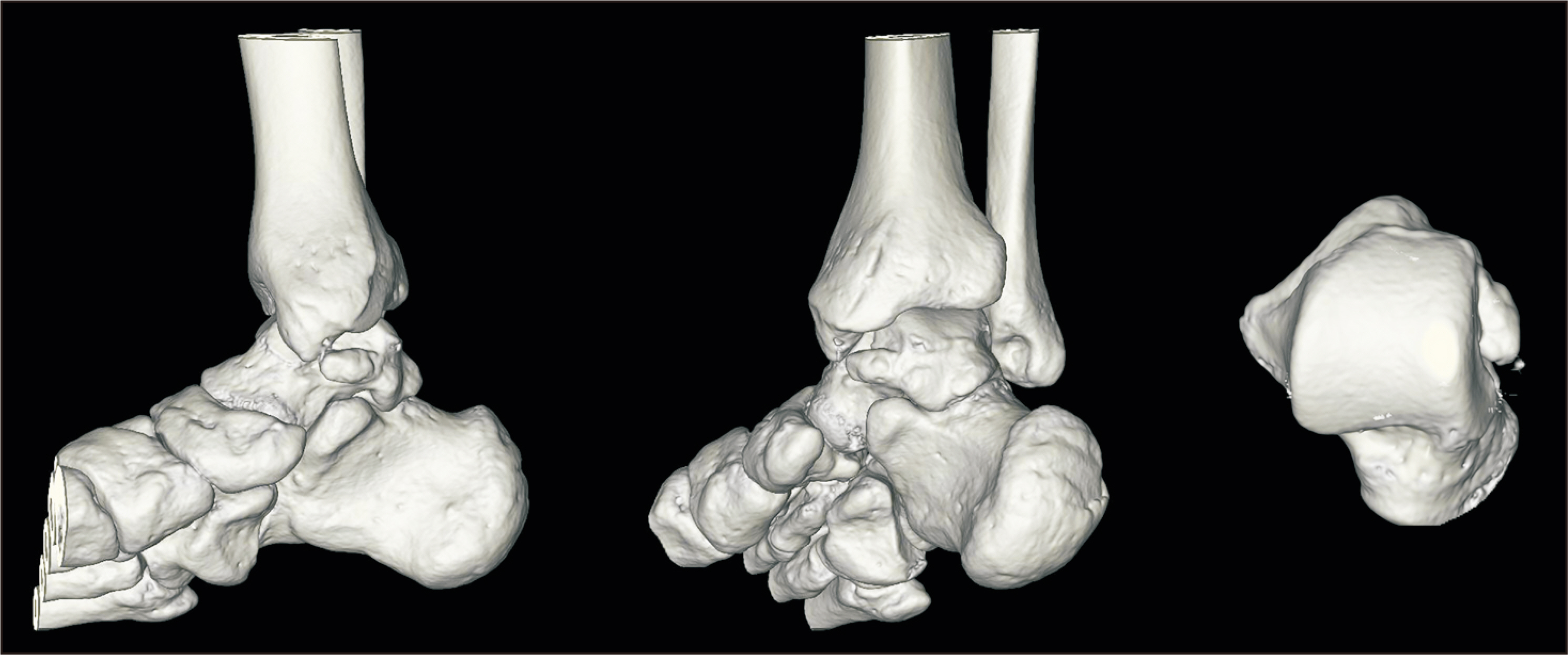
Figure 2 Computed tomography images showing an osteochondroma occurring on the medial tubercle of the talus.
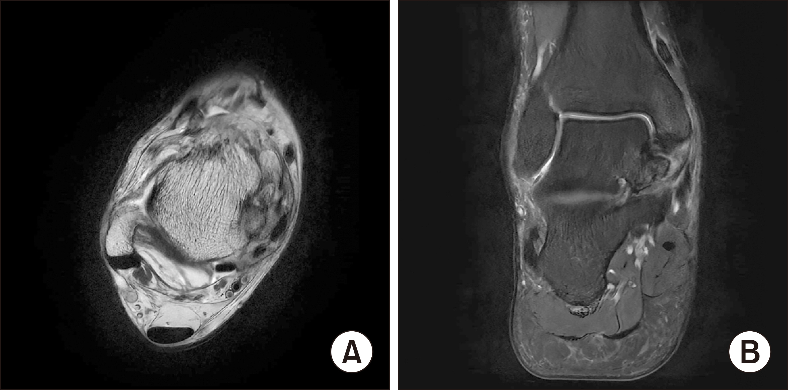
Figure 3 (A) T1- and (B) T2-weighted magnetic resonance images showing osteochondroma in the talus medial tubercle, a moderate amount of joint effusion with synovitis, and subcortical cysts in the medial malleolus.

Figure 4 Intraoperative medical photo. (A) Identification of the lesion (tip of the needle) by retracting the tibialis posterior tendon, (B) after complete excision of the lesion, (C) excised talar osteochondroma, and (D) gout tophi.
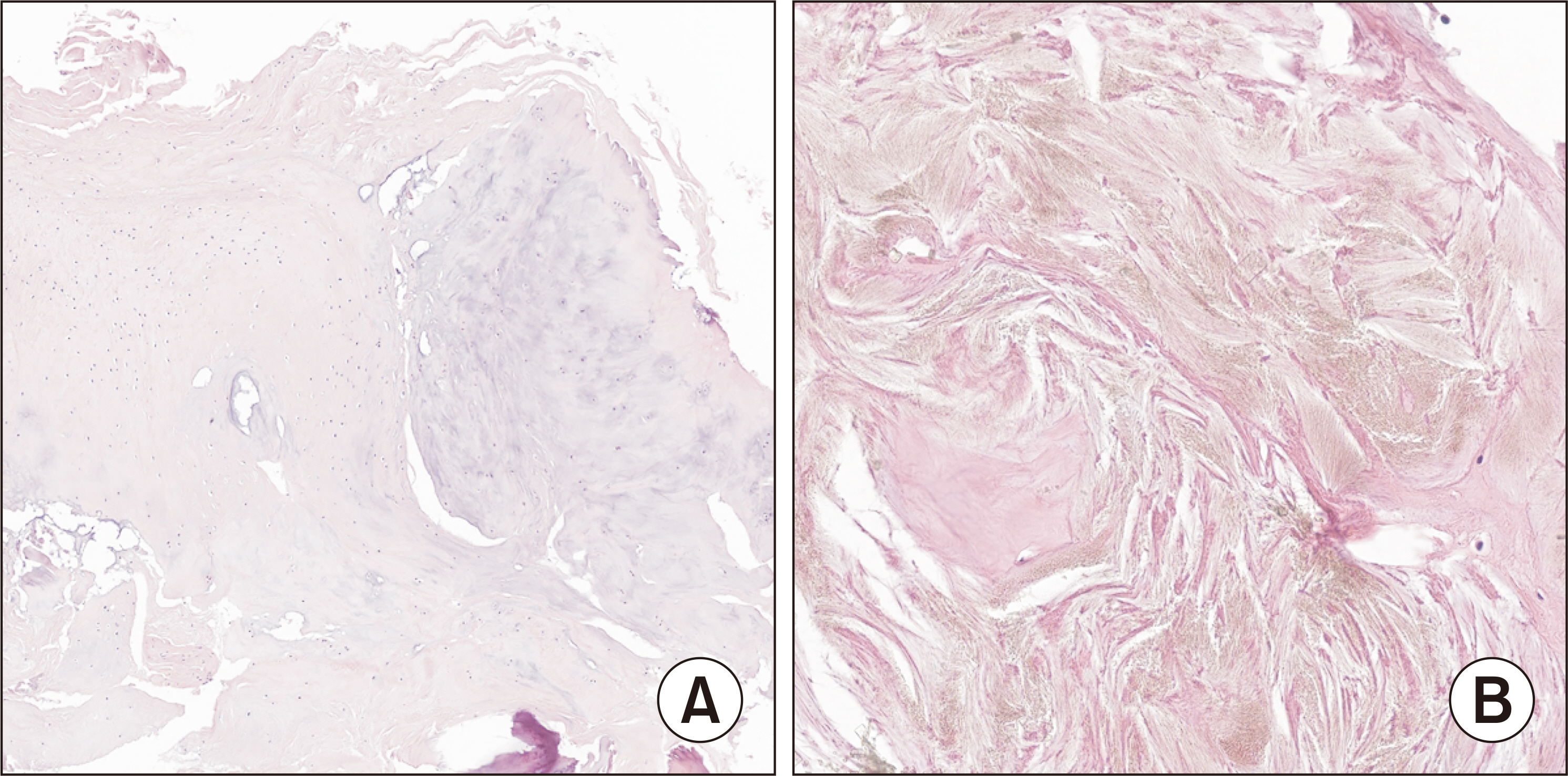
Figure 5 Microscopic slide view of (A) osteochondroma with a cartilage cap (H&E, ×400) and (B) deposition of needle shaped crystals indicating gout tophi (H&E, ×1,600).
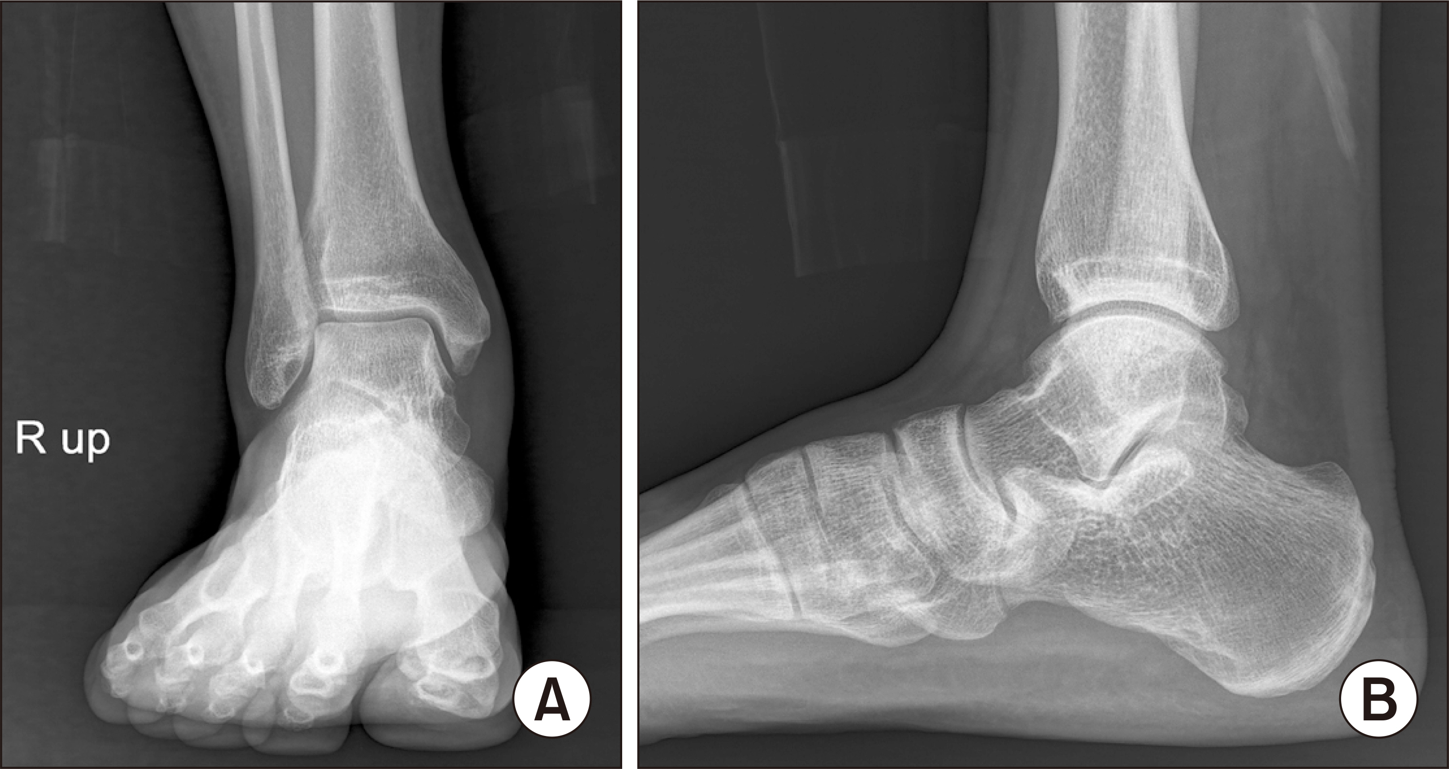
Figure 6 Final standing ankle (A) anteroposterior and (B) lateral radiographs 6 months after surgery showing no recurrence.
Reference
-
1. Wang C, Ma X, Wang X, Zhang Y, Zhang C, Huang J, et al. 2016; Osteochondroma of the talar neck: a case report and literature review. J Foot Ankle Surg. 55:338–44. doi: 10.1053/j.jfas.2014.10.005. DOI: 10.1053/j.jfas.2014.10.005. PMID: 25458440.
Article2. Galanis V, Georgiadi K, Balomenos V, Tsoucalas G, Thomaidis V, Fiska A. 2020; Osteochondroma of the talus in a 19-year-old female: a case report and review of the literature. Foot (Edinb). 42:101635. doi: 10.1016/j.foot.2019.08.006. DOI: 10.1016/j.foot.2019.08.006. PMID: 31739169.
Article3. Turan A, Kose O, Acar B, Unal M. 2017; Posterior tibial tendon impingement due to os subtibiale: a case report and up-to-date review. Skeletal Radiol. 46:705–14. doi: 10.1007/s00256-017-2601-1. DOI: 10.1007/s00256-017-2601-1. PMID: 28255943.
Article4. Kraus VB, Kilfoil TM, Hash TW 2nd, McDaniel G, Renner JB, Carrino JA, et al. 2015; Atlas of radiographic features of osteoarthritis of the ankle and hindfoot. Osteoarthritis Cartilage. 23:2059–85. doi: 10.1016/j.joca.2015.08.008. DOI: 10.1016/j.joca.2015.08.008. PMID: 26318654. PMCID: PMC4663119.
Article5. Kulkarni U, Kulkarni A. 2015; Posterior talus osteochondroma a rare location, treated by posterior ankle arthroscopy. Foot Ankle Surg. 21:e51–4. doi: 10.1016/j.fas.2015.05.011. DOI: 10.1016/j.fas.2015.05.011. PMID: 26235872.
Article6. Boya H, Ozcan O, Tokyol C. 2014; Osteochondroma of the talus: an unusual location. Acta Orthop Traumatol Turc. 48:236–9. doi: 10.3944/AOTT.2014.3217. DOI: 10.3944/AOTT.2014.3217. PMID: 24747637.
Article7. Shah A, Adhikari S, Shah S, Bhandari PS, Uprety S. 2022; Osteochondroma of talus: a case report from Nepal. Clin Case Rep. 10:e05867. doi: 10.1002/ccr3.5867. DOI: 10.1002/ccr3.5867. PMID: 35592046. PMCID: PMC9097038.
Article8. Joshi D, Kumar N, Singh D, Lal Y, Singh AK. 2005; Osteochondroma of the talus in a male adolescent. J Am Podiatr Med Assoc. 95:494–6. doi: 10.7547/0950494. DOI: 10.7547/0950494. PMID: 16166471.
Article9. Kumar S, Dhammi IK, Jain AK, Shahi P. 2020; Osteochondroma of the talus: three varying cases. BMJ Case Rep. 13:e237670. doi: 10.1136/bcr-2020-237670. DOI: 10.1136/bcr-2020-237670. PMID: 33370985. PMCID: PMC7757478.
Article10. Jackson KR, Gurbani B, Otsuka NY. 2004; Osteochondromas of the talus presenting as intraarticular loose bodies: report of two cases. Foot Ankle Int. 25:630–1. doi: 10.1177/107110070402500906. DOI: 10.1177/107110070402500906. PMID: 15563384.
Article11. Cavalcante MM, Silveira CRS, da Costa CR, Távora DGF, Alencar CHMF, Teixeira MJD, et al. 2021; Tumors and pseudotumors of foot and ankle: bone lesions. Foot (Edinb). 49:101845. doi: 10.1016/j.foot.2021.101845. DOI: 10.1016/j.foot.2021.101845. PMID: 34560430.
Article12. Mujaddid I, Rosihan E, Handry TH, Brian W. 2020; Tophaceous gout of hip joint mimicking bone tumor. Open Access Maced J Med Sci. 8(C):181–3. doi: 10.3889/oamjms.2020.4624. DOI: 10.3889/oamjms.2020.4624.
Article
