Multiple Aneurysms with Thrombosis on the Small Saphenous Vein
- Affiliations
-
- 1Department of Orthopedic Surgery, Chungnam National University School of Medicine, Daejeon, Korea
- 2Department of Orthopedic Surgery, Konyang University Hospital, Daejeon, Korea
- 3Department of Orthopedic Surgery, Yeonsei Barochuk Hospital, Seoul, Korea
- 4Department of Orthopedic Surgery, Keck School of Medicine of University of Southern California, CA, USA
- KMID: 2553806
- DOI: http://doi.org/10.14193/jkfas.2024.28.1.27
Abstract
- Several types of soft tissue masses occur in the lower extremities. A mass associated with blood vessels is often difficult to diagnose. A 15-year-old male patient visited the author’s hospital with discomfort and edema in his right calf that had persisted for six months. A physical examination showed no palpable mass other than mild edema. Three masses were found during the ultrasound scan along the small saphenous vein. The masses had a cyst-like appearance and were filled with thrombus. In duplex ultrasound, vascular reflux was represented inside the masses. During surgery, it was suspected that vascular deformation occurred in the small saphenous vein, and simple ligation and resection treatments were performed. The patient was finally diagnosed with venous aneurysms accompanied by thrombosis based on the histology tests. The symptoms disappeared after surgery, and there were no recurrences or unusual findings at the follow-up one year later. Venous aneurysms occurring in the superficial veins of the lower extremities are rarely reported, but treatment and diagnosis are important. This paper reports a case of an aneurysm on the small saphenous vein.
Keyword
Figure
-
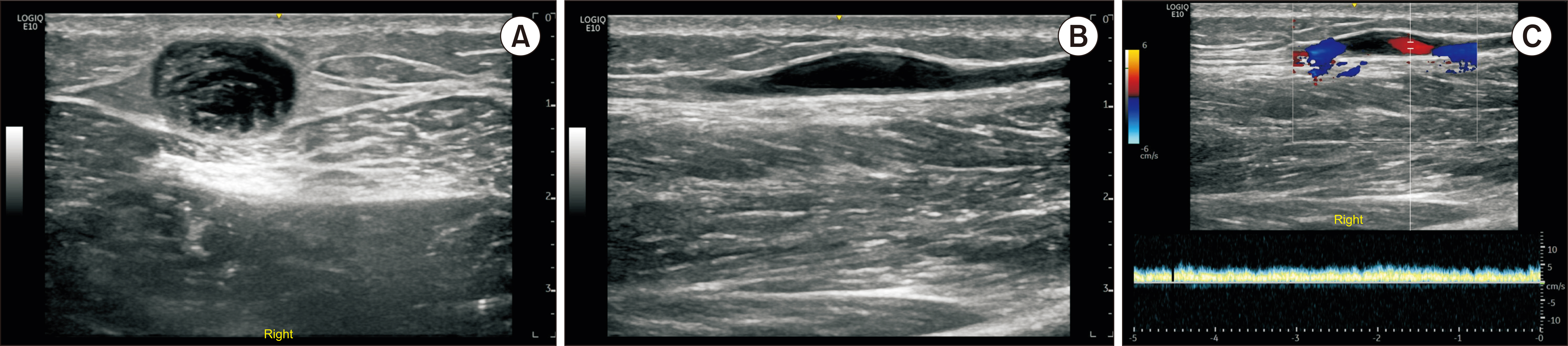
Figure 1 The ultrasound images of the right small saphenous vein. (A) In the short axis view, the vein is dilated as fusiform shape aneurysm containing thrombus. (B) In the long axis view, blood vessel is dilated about twice as much as the normal areas above and below. (C) Duplex ultrasound scan showing venous reflux in the aneurysm.

Figure 2 The magnetic resonance imaging images. (A) T1-weighted axial, (B) T2-weighted longitudinal, (C, D) T1-weighted enhanced axial and longitudinal images show the dilatation and thrombus along the small saphenous vein.

Figure 3 The gross photo images. (A) Venous aneurysms are marked on the posterior calf by using ultrasound in the operation room. (B) These tissues are the proximal (arrow) and distal (arrowhead) aneurysms after aneurysmectomy.
Reference
-
1. Kransdorf MJ, Murphey MD. 2006. Imaging of soft tissue tumors. 2nd ed. Lippincott Williams & Wilkins;Philadelphia:2. Gabrielli R, Rosati MS, Siani A, Irace L. 2012; Management of symptomatic venous aneurysm. ScientificWorldJournal. 2012:386478. doi: 10.1100/2012/386478. DOI: 10.1100/2012/386478. PMID: 22566766. PMCID: PMC3329879.
Article3. Pascarella L, Al-Tuwaijri M, Bergan JJ, Mekenas LM. 2005; Lower extremity superficial venous aneurysms. Ann Vasc Surg. 19:69–73. doi: 10.1007/s10016-004-0135-1. DOI: 10.1007/s10016-004-0135-1. PMID: 15714370.
Article4. Maldonado-Fernandez N, Lopez-Espada C, Martinez-Gamez FJ, Galan-Zafra M, Sanchez-Maestre ML, Herrero-Martinez E, et al. 2013; Popliteal venous aneurysms: results of surgical treatment. Ann Vasc Surg. 27:501–9. doi: 10.1016/j.avsg.2012.07.005. DOI: 10.1016/j.avsg.2012.07.005. PMID: 23522443.
Article5. Sessa C, Nicolini P, Perrin M, Farah I, Magne JL, Guidicelli H. 2000; Management of symptomatic and asymptomatic popliteal venous aneurysms: a retrospective analysis of 25 patients and review of the literature. J Vasc Surg. 32:902–12. doi: 10.1067/mva.2000.110353. DOI: 10.1067/mva.2000.110353. PMID: 11054222.
Article6. Irwin C, Synn A, Kraiss L, Zhang Q, Griffen MM, Hunter GC. 2008; Metalloproteinase expression in venous aneurysms. J Vasc Surg. 48:1278–85. doi: 10.1016/j.jvs.2008.06.056. DOI: 10.1016/j.jvs.2008.06.056. PMID: 18971037.
Article7. Aggarwal V. 2021; Pathogenesis and management of superficial venous aneurysms through a case of thrombosed large great saphenous vein aneurysm. Vascular. 29:297–300. doi: 10.1177/1708538120949720. DOI: 10.1177/1708538120949720. PMID: 32829698.
Article8. Lee HY, Lee W, Cho YK, Chung JW, Park JH. 2006; Superficial venous aneurysm: reports of 3 cases and literature review. J Ultrasound Med. 25:771–6. doi: 10.7863/jum.2006.25.6.771. DOI: 10.7863/jum.2006.25.6.771. PMID: 16731894.9. Bergqvist D, Björck M, Ljungman C. 2006; Popliteal venous aneurysm--a systematic review. World J Surg. 30:273–9. doi: 10.1007/s00268-005-7982-y. DOI: 10.1007/s00268-005-7982-y. PMID: 16479328.
Article10. Hallstensson S, Ljungman C, Rudström H, Björck M, Bergqvist D. 2005; [Claudication and pulmonary embolism can be caused by venous aneurysm. A case report illustrates difficulties with this unusual diagnosis]. Lakartidningen. 102:1152–3. Swedish.11. Gillespie DL, Villavicencio JL, Gallagher C, Chang A, Hamelink JK, Fiala LA, et al. 1997; Presentation and management of venous aneurysms. J Vasc Surg. 26:845–52. doi: 10.1016/s0741-5214(97)70099-5. DOI: 10.1016/S0741-5214(97)70099-5. PMID: 9372824.
Article
- Full Text Links
-
- Actions
-
Cited
- CITED
-
- Close
- Share
-
- Similar articles
-
- Prevalence and Clinical Implication of Nonsaphenous Vein Reflux with or without Pelvic Venous Disease
- An Acute Pulmonary Embolism Accompanying Greater Saphenous Vein Thrombosis
- Endovenous Laser Treatment (EVLT) with High Ligation of an Incompetent Small Saphenous Vein
- Effect of Diameter of Saphenous Vein on Stump Length after Radiofrequency Ablation for Varicose Vein
- Patterns of Saphenous Vein Reflux and Treatment Plan
