Arch Hand Microsurg.
2024 Mar;29(1):60-63. 10.12790/ahm.23.0053.
Painful eccrine spiradenoma containing nerve fibers in the forearm: a case report
- Affiliations
-
- 1Department of Plastic and Reconstructive Surgery, CHA Bundang Medical Center, CHA University School of Medicine, Seongnam, Korea
- 2April 31 Plastic Surgery Clinic, Seoul, Korea
- KMID: 2553173
- DOI: http://doi.org/10.12790/ahm.23.0053
Abstract
- Eccrine spiradenomas are rare benign adnexal tumors that originate from the eccrine glands. They mainly arise in the head, neck, and upper trunk of young adults and are sometimes accompanied by pain and tenderness. Although spontaneous pain is a typical symptom of eccrine spiradenoma, the underlying mechanism remains unclear. We report the case of a patient who had a spiradenoma in the subcutaneous tissue of the left forearm that was accompanied by agonizing pain triggered by pressure. A 42-year-old man who had undergone surgical excision of a left forearm mass 5 years ago presented for relapsed pain and a palpable mass at the previous excision site. The agonizing pain had been triggered a few days ago, in response to even a slight touch. The mass measured approximately 0.8×0.8 mm, and ther e were no changes visible around the scar. Irregularly shaped masses, each measuring approximately 5 mm, were removed with a 2-mm safety margin from the subcutaneous fat. A histopathologic examination revealed the resected nodules were surrounded by delicate fibrous capsules, some parts of which contained blood vessels and prominent thickened nerve fibers. By the time of the 6-month follow-up, the masses had not recurred, and the biopsy site was well maintained without pain. We describe the surgical and histopathologic findings of an isolated eccrine spiradenoma accompanied by recurrent agonizing pain. The peculiar microscopic arrangement of the thickened nerve fibers encasing the nodule of eccrine spiradenoma may correlate with its pain mechanism.
Figure
-

Fig. 1. A solitary mass on the left forearm of a 42-year-old man.
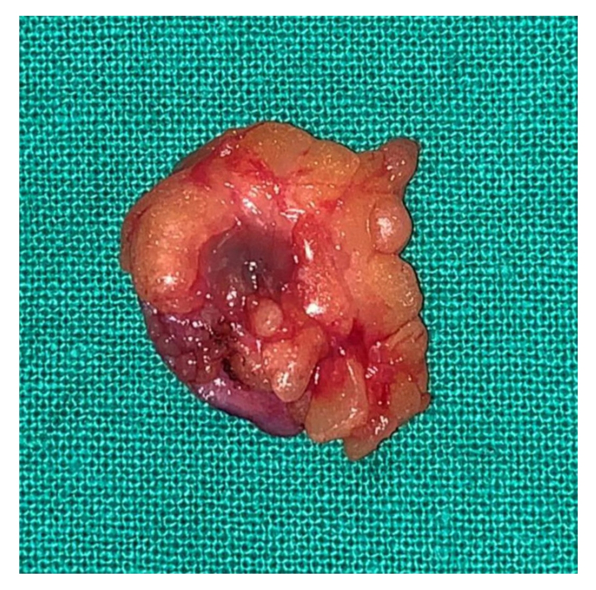
Fig. 2. The resected specimen with fat tissue, approximately 5 mm in size.
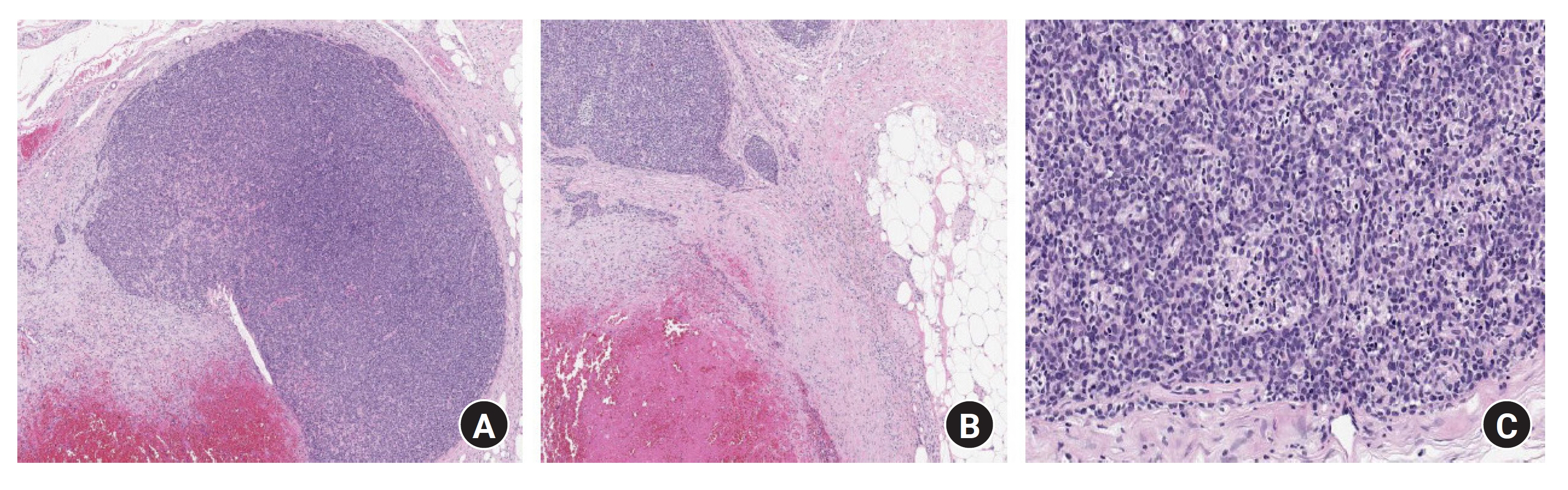
Fig. 3. Microscopic findings (hematoxylin and eosin staining). (A) Well-circumscribed and encapsulated tumor nodule with smooth borders in the dermis and subcutaneous tissue, some of which contained blood vessels and prominent thickened nerve fibers (×40). (B) A capsule area containing nerve fibers (x40). (C) Tumor lobules composed of two cell types. Peripherally, smaller dark blue cells and centrally, pale larger cells. (x100).
Reference
-
References
1. Kersting DW, Helwig EB. Eccrine spiradenoma. AMA Arch Derm. 1956; 73:199–227.
Article2. Naversen DN, Trask DM, Watson FH, Burket JM. Painful tumors of the skin: "LEND AN EGG". J Am Acad Dermatol. 1993; 28(2 Pt 2):298–300.
Article3. Syauqi MS, Marliza H, BHK NG. A rare adnexal tumor of head & neck: eccrine spiradenoma. J Dermatol Res Therapy. 2021; 7:102.
Article4. Byeon JY, Kim JH. Solitary spiradenoma of the hand misdiagnosed as neurilemmoma. Arch Hand Microsurg. 2018; 23:190–4.
Article5. Kim J, Yang HJ, Pyo JS. Eccrine spiradenoma of the scalp. Arch Craniofac Surg. 2017; 18:211–3.
Article6. Ohtsuki Y, Ohtsuka H, Kurabayashi A, et al. Immunohistochemical and electron microscopic studies of Langerhans cells in a case of multiple eccrine spiradenomas. Med Mol Morphol. 2007; 40:221–5.
Article7. Mambo NC. Eccrine spiradenoma: clinical and pathologic study of 49 tumors. J Cutan Pathol. 1983; 10:312–20.
Article8. Park HR, Im SB, Kim HK, Shin DS, Park YL. Painful eccrine spiradenoma containing nerve fibers: a case report. Dermatology. 2012; 224:301–6.
Article9. Kolda TF, Ardaman TD, Schwartz MR. Eccrine spiradenoma mimicking adenoid cystic carcinoma on fine needle aspiration. A case report. Acta Cytol. 1997; 41:852–8.10. Castro C, Winkelmann RK. Spiradenoma. Histochemical and electron microscopic study. Arch Dermatol. 1974; 109:40–8.
Article
