Ann Lab Med.
2023 Mar;43(2):208-210. 10.3343/alm.2023.43.2.208.
Whole-mount Electron Microscopy to Quantitate Platelet Dense Granules: Reference Intervals for Healthy Controls in Korea
- Affiliations
-
- 1Department of Laboratory Medicine, Dong-A University College of Medicine, Busan, Korea
- 2Department of Laboratory Medicine, Dong-A University Hospital, Busan, Korea
- 3Department of Pathology, Dong-A University College of Medicine, Busan, Korea
- KMID: 2551700
- DOI: http://doi.org/10.3343/alm.2023.43.2.208
Figure
-
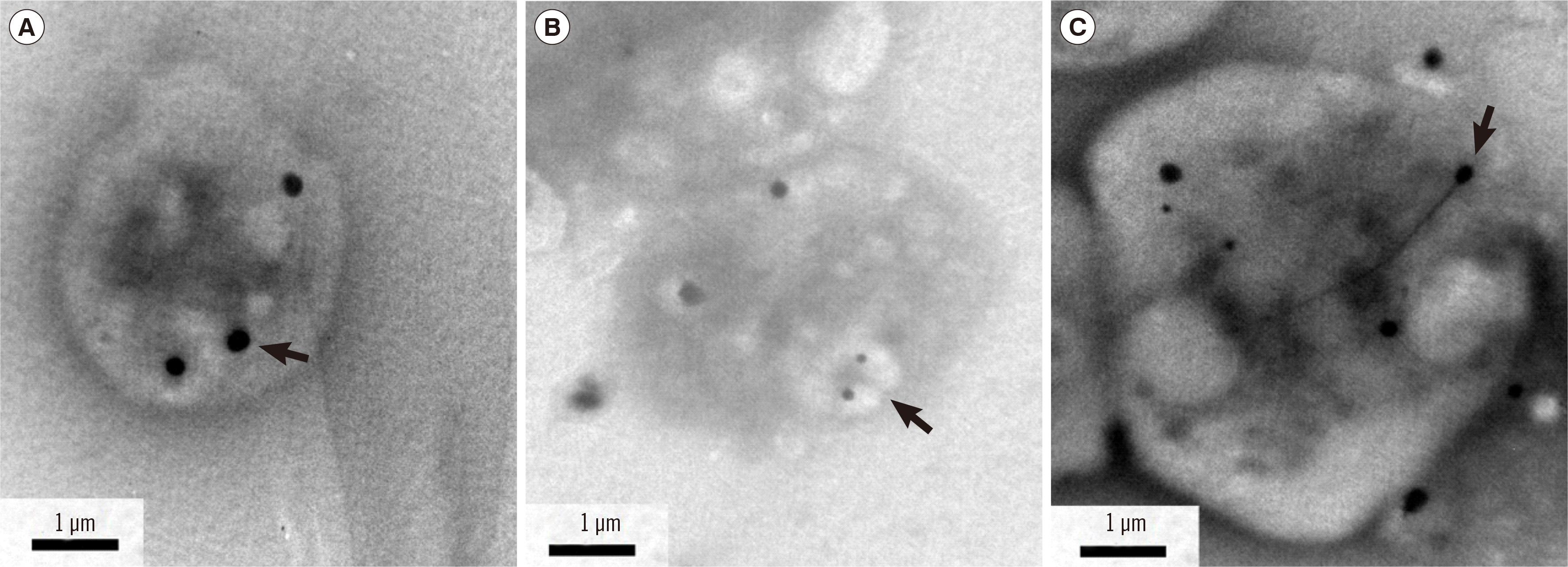
Fig. 1 Representative electron micrographs of platelet whole-mount preparations from healthy individuals. (A) Electron-opaque dense bodies. (B) Granules enclosed by membranes in hyaloplasm. (C) Dense granules with long tails (arrows, ×12,000 magnification).

Fig. 2 Dense granule counts in platelets from healthy adults. Box plots showing midspreads and total ranges in women and men. The vertical axis indicates the number of dense granules per platelet.
Reference
-
1. Hayward CP, Moffat KA, Plumhoff E, Van Cott EM. Approaches to investigating common bleeding disorders: an evaluation of North American coagulation laboratory practices. Am J Hematol. 2012; 87(S1):S45–50. DOI: 10.1002/ajh.23124. PMID: 22367923.
Article2. Brunet JG, Iyer JK, Badin MS, Graf L, Moffat KA, Timleck M, et al. 2018; Electron microscopy examination of platelet whole mount preparations to quantitate platelet dense granule numbers: implications for diagnosing suspected platelet function disorders due to dense granule deficiency. Int J Lab Hematol. 40:400–7. DOI: 10.1111/ijlh.12801. PMID: 29508516.
Article3. Chen Y, Yuan Y, Li W. 2018; Sorting machineries: how platelet-dense granules differ from α-granules. Biosci Rep. 38:BSR20180458. DOI: 10.1042/BSR20180458. PMID: 30104399. PMCID: PMC6127676.
Article4. Sorokin V, Alkhoury R, Al-Rawabdeh S, Houston RH, Thornton D, Kerlin B, et al. 2016; Reference range of platelet delta granules in the pediatric age group: an ultrastructural study of platelet whole mount preparations from healthy volunteers. Pediatr Dev Pathol. 19:498–501. DOI: 10.2350/15-11-1733-OA.1. PMID: 26670096.
Article5. Hayward CP, Moffat KA, Spitzer E, Timleck M, Plumhoff E, Israels SJ, et al. 2009; Results of an external proficiency testing exercise on platelet dense-granule deficiency testing by whole mount electron microscopy. Am J Clin Pathol. 131:671–5. DOI: 10.1309/AJCPYMFXZNSNRZ55. PMID: 19369626.
Article6. Dupuis A, Bordet JC, Eckly A, Gachet C. 2020; Platelet δ-storage pool disease: an update. J Clin Med. 9:2508. DOI: 10.3390/jcm9082508. PMID: 32759727. PMCID: PMC7466064.
Article7. Gunning WT 3rd, Raghavan M, Calomeni EP, Turner JN, Roysam B, Roysam S, et al. 2020; A morphometric analysis of platelet dense granules of patients with unexplained bleeding: a new entity of delta-microgranular storage pool deficiency. J Clin Med. 9:1734. DOI: 10.3390/jcm9061734. PMID: 32512725. PMCID: PMC7356033.
Article8. Urban D, Pluthero FG, Christensen H, Baidya S, Rand ML, Das A, et al. 2017; Decreased numbers of dense granules in fetal and neonatal platelets. Haematologica. 102:e36–8. DOI: 10.3324/haematol.2016.152421. PMID: 27810994. PMCID: PMC5286950.
Article9. Tian J, Cheng LH, Cui X, Lei XX, Tang JB, Cheng B. 2019; Investigating the effect of age on platelet ultrastructure using transmission electron microscopy. Int Wound J. 16:1457–63. DOI: 10.1111/iwj.13214. PMID: 31486290. PMCID: PMC7949169.
Article
- Full Text Links
-
- Actions
-
Cited
- CITED
-
- Close
- Share
-
- Similar articles
-
- The Ultrastructure of Tongue Mast Cells in Bombina orientalis
- Ultrastructural Study of Crypt Cells in the Ileum of Mouse after Salmonella typhimurium Infection
- Ultrastructural Changes of the Rabbit Lens Fiber Cells in Cataract
- Transmission electron microscopic findings of the tribocytic organ of Fibricola seoulensis
- A Case of Congenital Self-Healing Reticulohistiocytosis
