Guidewire insertion into the vertebral vein during right internal jugular vein central venous catheterization -A rare case report-
- Affiliations
-
- 1Department of Anesthesiology and Pain Medicine, Inje University Busan Paik Hospital, Inje University College of Medicine, Busan, Korea
- KMID: 2550918
- DOI: http://doi.org/10.17085/apm.23052
Abstract
- Background
Internal jugular veins are the most frequently accessed site for central venous catheterization in patient management, whereas complications involving vertebral veins are a rare occurrence. Case: A 73-year-old male suspected to have a urothelial carcinoma was scheduled for elective left nephroureterectomy. During central venous catheterization using the anatomic landmark technique to target the internal jugular vein, a guidewire is inadvertently inserted into the suspected vertebral vein. Following the correction of the catheterization, a radiologist reviewed the preoperative enhanced computed tomography and confirmed that the initially punctured vessel was the vertebral vein. On the third day after surgery, the central venous catheter was removed, and the patient did not exhibit any complications, such as bleeding, swelling, and neurological symptoms. Conclusions: The use of ultrasonography during central venous catheterization is recommended to evaluate the anatomy of the puncture site and prevent misinsertion of the catheter, which can lead to several complications.
Keyword
Figure
-
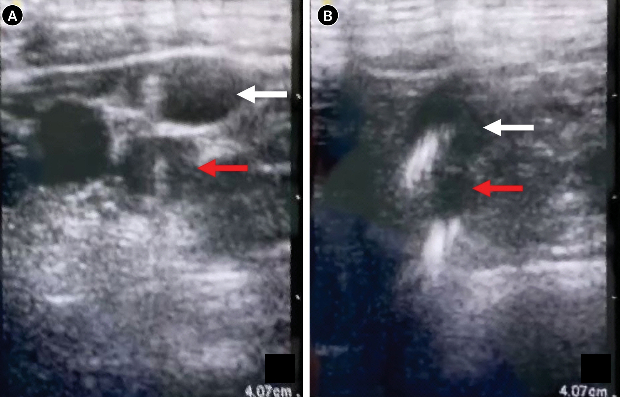
Fig. 1. Ultrasonography. (A) A larger vessel where the second guidewire was inserted is located superiorly and more laterally than the vessel that was first punctured. (B) The course of both veins, where the guidewires were inserted, descends toward the right brachiocephalic vein. Red arrow: vertebral vein; white arrow: internal jugular vein.
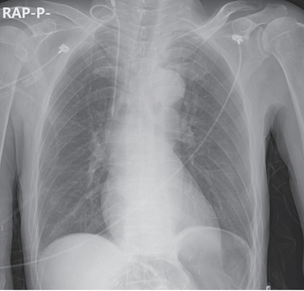
Fig. 2. The central venous catheter, which was cannulated using the second inserted guidewire, is located near the right atrium and follows the superior vena cava.
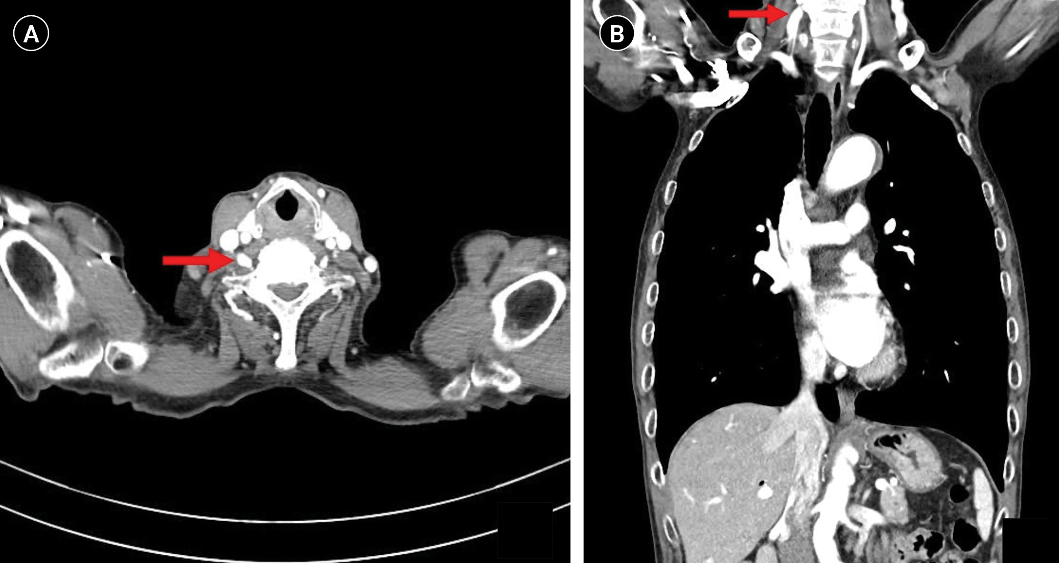
Fig. 3. Contrast-enhanced computed tomogram. (A) The axial view at the seventh cervical vertebral level reveals a suspected vein running from the vertebral foramen of the sixth cervical vertebra. (B) Coronary view of the same computed tomogram also reveals an escape of the suspected vein from the sixth vertebral foramen. Red arrow: suspected vein.
Reference
-
1. Turan S. Inadvertent vertebral vein catheterisation during transjugular vein cannulation: a rare complication. Turk Gogus Kalp Dama. 2013; 21:776–8.
Article2. Wang L, Liu ZS, Wang CA. Malposition of central venous catheter: presentation and management. Chin Med J (Engl). 2016; 129:227–34.3. Bannon MP, Heller SF, Rivera M. Anatomic considerations for central venous cannulation. Risk Manag Healthc Policy. 2011; 4:27–39.
Article4. M Bawa C, Dabla L, Raina J, Chadha H. Malpositioning of central venous catheter from right internal jugular vein into ipsilateral vertebral vein: a rare phenomenon. Indian J Public Health. 2020; 11:519–22.5. Yang SH, Jung SM, Park SJ. Misinsertion of central venous catheter into the suspected vertebral vein: a case report. Korean J Anesthesiol. 2014; 67:342–5.
Article6. Kobayashi Y, Hamano R, Watanabe H, Hong J, Toyoda K, Hashizume M, et al. Use of puncture force measurement to investigate the conditions of blood vessel needle insertion. Med Eng Phys. 2013; 35:684–9.
Article7. Kobayashi Y, Hong J, Hamano R, Okada K, Fujie MG, Hashizume M. Development of a needle insertion manipulator for central venous catheterization: needle insertion manipulator for central venous catheterization. Int J Med Robot. 2012; 8:34–44.
Article8. Ciuti G, Righi D, Forzoni L, Fabbri A, Pignone AM. Differences between internal jugular vein and vertebral vein flow examined in real time with the use of multigate ultrasound color doppler. AJNR Am J Neuroradiol. 2013; 34:2000–4.
Article9. Winston CB, Wechsler RJ, Kane M. Vertebral vein migration of a long-term central venous access catheter: a cause of brachial plexopathy. J Thorac Imaging. 1994; 9:98–100.10. Kulkarni A, Chandrakar N, Deokar S, Bhasin N. Accidental vertebral vein catheterization during internal jugular vein cannulation–A rare complication. J Anaesthesiol Clin Pharmacol. 2019; 35:277–8.
Article
- Full Text Links
-
- Actions
-
Cited
- CITED
-
- Close
- Share
-
- Similar articles
-
- Malposition of a Subclavian Catheter in the Internal Jugular Vein Due to the Direction of a J-type Guidewire End
- Knotting of a guidewire during internal jugular vein catheterization in an infant: A case report
- Guidewire Entrapment During Central Venous Catheterization
- Internal jugular vein thrombosis detection by ultrasound scan after failure of internal jugular vein catheterization: A case report
- Incidental ipsilateral subclavian vein catheterization via right internal jugular venous route: A case report
