Multiple fixed implant-supported prosthesis using temporary denture and scannable healing abutment: a case report
- Affiliations
-
- 1Department of Prosthodontics, School of Dentistry, Chonnam National University, Gwangju, Republic of Korea
- KMID: 2550434
- DOI: http://doi.org/10.14368/jdras.2023.39.4.250
Abstract
- The use of digital technology in fixed prosthetic treatment using implants enables predictive treatment through diagnosis and virtual surgery by integrating clinical and radiological information of patients. Existing digital scanning methods require several components to be removed, such as removing the healing abutment and connecting the scan body. In the scannable healing abutment developed in consideration of this point, scanning is performed directly on the healing abutment, maintaining soft tissue sealing and simplifying scanning. Digital technology can also be used when obtaining the intermaxillary relationship. Recently, various digital technologies have been reported to acquire the intermaxillary relationship of edentulous patients using surgical guides, patient-specific scanning devices, or scans of the inside of temporary dentures. In this case, the implant-supported fixed prosthesis treatment was performed through scanning the scannable healing abutment and the inner side of the temporary denture to obtain the intermaxillary relationship, thereby simplifying the treatment process and obtaining aesthetically and functionally excellent clinical results.
Figure
-

Fig. 1 Initial intraoral photographs. (A) Initial panoramic radiography, (B) Maxillary occlusal view, (C) Frontal view, (D) Mandibular occlusal view.

Fig. 2 Implant planning using R2GATE software. (A) Radiopaque occlusal rim, (B) Digital wax up, (C) Planned implant position of maxilla, (D) Planned implant position of mandible, (E) 3D printing surgical guide of maxilla, (F) 3D printing surgical guide of mandible.

Fig. 3 Mandibular anterior tooth state with Intraoral photo. (A) Frontal view, (B) Occlusal view, (C) Periapical radiography.

Fig. 4 Post-operative images after implant stage surgery. (A) Panoramic radiograph with healing scan abutment, (B) Occlusal view of maxilla with healing scan abutment, (C) Occlusal view of mandible with healing scan abutment.

Fig. 5 Temporary denture relining with tissue conditioner. (A) Frontal view, Inner surface of temporary denture of (B) Maxilla, (C) Mandible.

Fig. 6 Superimposition between temporary denture scan data and intraoral scan data. Intraoral scan data of (A) Maxilla, (B) Mandible. Temporary denture scan data of (C) Maxilla, (D) Mandible and Inverted scan data of (E) Maxilla, (F) Mandible. (G) Frontal view, (H) Sagittal view of alignment of scan data with facial scan data and evaluation of occlusal plane.

Fig. 7 Design of provisional prosthesis with superimposition data by Exocad. (A) Frontal view, (B) Sagittal view.
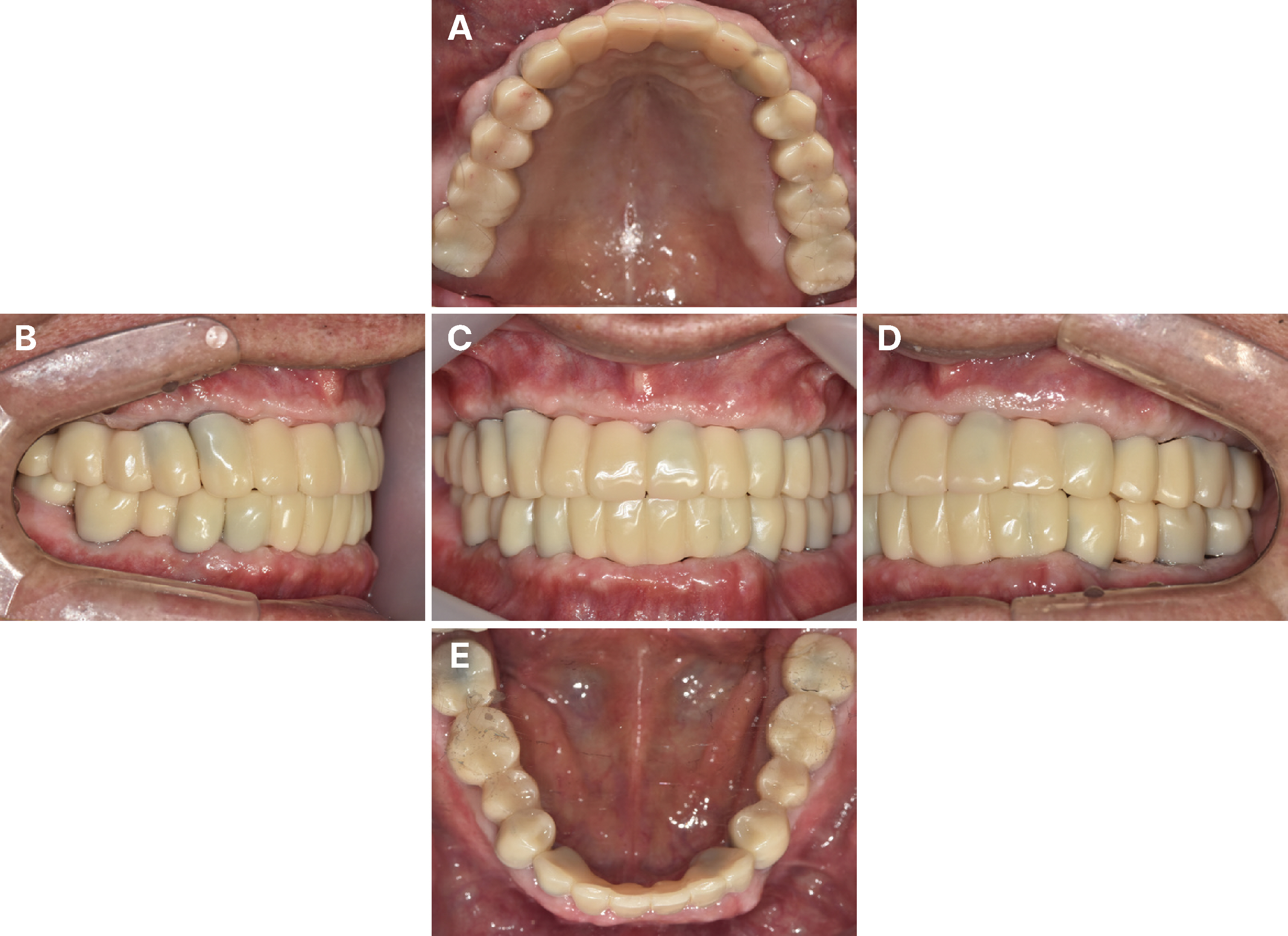
Fig. 8 Provisional prosthesis. (A) Occlusal view of maxilla, (B) Right lateral view, (C) Fontal view, (D) Left lateral view, (E) Occlusal view of mandible.

Fig. 9 (A) Bite taking with bite impression material, Abutment level impression of (B) Maxiila, (C) Mandible, (D - F) Cross mounting for definitive prosthesis, (G, H) Mandibular movement record with Arcus digma.

Fig. 10 Delivery of definitive prosthesis (Posterior part). (A) Occlusal view of maxilla, (B) Right lateral view, (C) Fontal view, (D) Left lateral view, (E) Occlusal view of mandible, (F) Extraoral photograph (initial visit), (G) Extraoral photograph (after definitive prosthesis delivery).
Reference
-
References
1. Ender A, Mehl A. 2015; In-vitro evaluation of the accuracy of conventional and digital methods of obtaining full-arch dental impressions. Quintessence Int. 46:9–17. DOI: 10.3290/j.qi.a32244. PMID: 25019118.2. Alghazzawi TF. 2016; Advancements in CAD/CAM technology: Options for practical implementation. J Prosthodont Res. 60:72–84. DOI: 10.1016/j.jpor.2016.01.003. PMID: 26935333.3. Atsuta I, Ayukawa Y, Kondo R, Oshiro W, Matsuura Y, Furuhashi A, Tsukiyama Y, Koyano K. 2016; Soft tissue sealing around dental implants based on histological interpretation. J Prosthodont Res. 60:3–11. DOI: 10.1016/j.jpor.2015.07.001. PMID: 26725967.4. Krahenbuhl JT, Cho SH, Irelan J, Bansal NK. 2016; Accuracy and precision of occlusal contacts of stereolithographic casts mounted by digital interocclusal registrations. J Prosthet Dent. 116:231–6. DOI: 10.1016/j.prosdent.2016.01.029. PMID: 27068319.5. Ahmed WM, Verhaeghe TV, McCullagh APG. 2021; Maxillary complete-arch implant-supported restoration: a digital scanning and maxillomandibular relationship workflow. J Prosthet Dent. 125:216–20. DOI: 10.1016/j.prosdent.2020.01.010. PMID: 32171489.6. Ramadan RE, Bahgat MM, Abdelhamid AM, Khamis MM. 2023; Jan. 4. Registration of maxillomandibular relationship through a fully digital workflow for complete-mouth rehabilitation with screw-retained fixed implant-supported prostheses: A clinical report. J Prosthet Dent. S0022-3913(22)00760-0. DOI: 10.1016/j.prosdent.2022.11.027. PMID: 36609083.7. Eliasson A, Ortorp A. 2012; The accuracy of an implant impression technique using digitally coded healing abutments. Clin Implant Dent Relat Res. 14 Suppl 1:e30–8. DOI: 10.1111/j.1708-8208.2011.00344.x. PMID: 21453396.8. Ahn GZ, Lee JS. 2020; Comparison of the accuracy of implant digital impression coping. J Dent Rehabil Appl Sci. 36:29–40. DOI: 10.14368/jdras.2020.36.1.29.9. Jung HT, Kim HY, Song SY, Park JH, Lee JY. 2022; Accuracy of implant impression techniques with a scannable healing abutment. J Prosthet Dent. 128:729–34. DOI: 10.1016/j.prosdent.2020.06.042. PMID: 33832762.10. Abdulmajeed AA, Lim KG, Närhi TO, Cooper LF. 2016; Complete-arch implant-supported monolithic zirconia fixed dental prostheses: A systematic review. J Prosthet Dent. 115:672–7. DOI: 10.1016/j.prosdent.2015.08.025. PMID: 26809220.11. Joda T, Gallucci GO. 2015; The virtual patient in dental medicine. Clin Oral Implants Res. 26:725–26. DOI: 10.1111/clr.12379. PMID: 24665872.12. Kim JE, Park JH, Moon HS, Shim JS. 2019; Complete assessment of occlusal dynamics and establishment of a digital workflow by using target tracking with a three-dimensional facial scanner. J Prosthodont Res. 63:120–4. DOI: 10.1016/j.jpor.2018.10.003. PMID: 30446410.
- Full Text Links
-
- Actions
-
Cited
- CITED
-
- Close
- Share
-
- Similar articles
-
- Posterior single implant prosthesis using scannable healing abutment
- Full mouth rehabilitation with fixed implant-supported prosthesis using temporary denture and double digital scanning technique: a case report
- Maxillary implant-supported overdenture with magnetic attachment using healing abutment: A case report
- Implant-assisted removable partial denture using freely removable abutment in a fully edentulous patient: A case report
- Removable Partial Denture Using Anterior Implant-Supported Fixed Prostheses for Edentulous Patients: A Case Report
