Fabrication of implant-associated obturator after extraction of abutment teeth: a case report
- Affiliations
-
- 1Department of Prosthodontics, College of Dentistry, Chosun University, Gwangju, Republic of Korea
- KMID: 2550432
- DOI: http://doi.org/10.14368/jdras.2023.39.4.229
Abstract
- Maxillary bone defects may follow surgical treatment of benign and malignant tumors, trauma, and infection. Palatal defects often lead to problems with swallowing and pronunciation from the leakage of air into the nasal cavity and sinus. Obturators have been commonly used to solve these problems, but long-term use of the device may cause irritation of the oral mucosa or damage to the abutment teeth. Utilizing implants in the edentulous area for the fabrication of the obturators has gained attention. This case report describes a patient, who had undergone partial resection of the maxilla due to adenocarcinoma, in need of a new obturator after losing abutment teeth after long-term use of the previous obturator. Implants were placed in strategic locations, and an implantretained maxillary obturator was fabricated, showing satisfactory results in the rehabilitation of multiple aspects, including palatal defect, masticatory function, swallowing, pronunciation, and aesthetics.
Keyword
Figure
-
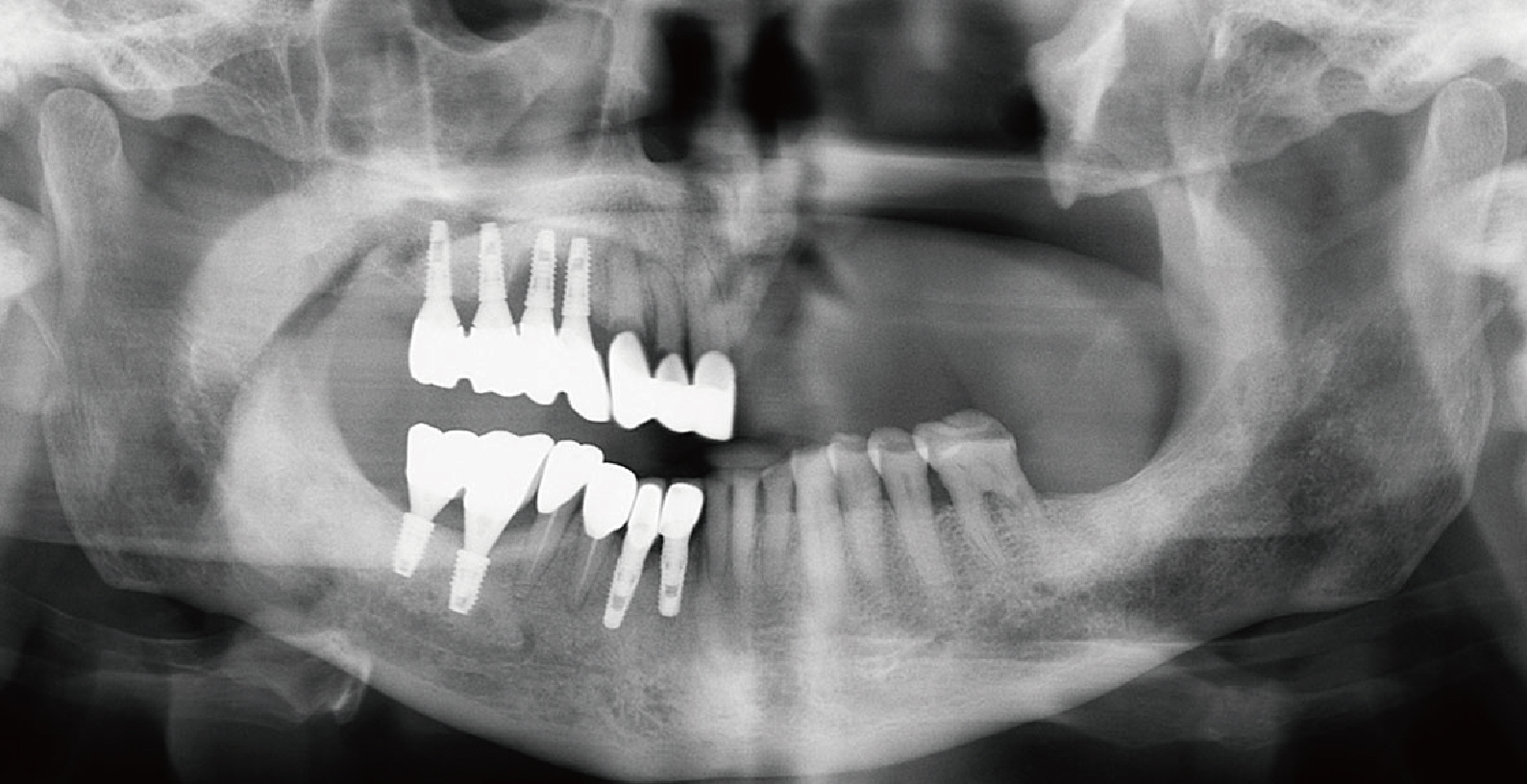
Fig. 1 Panoramic radiograph before treatment. Maxillary lateral incisor and canine have severe secondary caries and mobility.
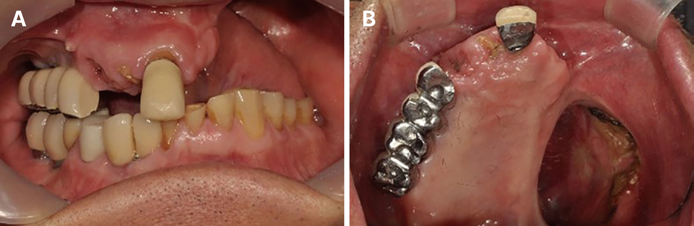
Fig. 2 Intraoral photograph before treatment. Rt. maxillary lateral incisor and canine were fractured due to severe secondary caries. Bridge of Rt. maxillary lateral incisor and canine was removed. (A) Frontal view, (B) Occlusal view.

Fig. 3 Intraoral photograph and old obturator photograph. Minor connector at Rt. maxillary first premolar and second premolar was fractured (Red circle). (A) Occlusal view, (B) Tissue surface of old obturator.
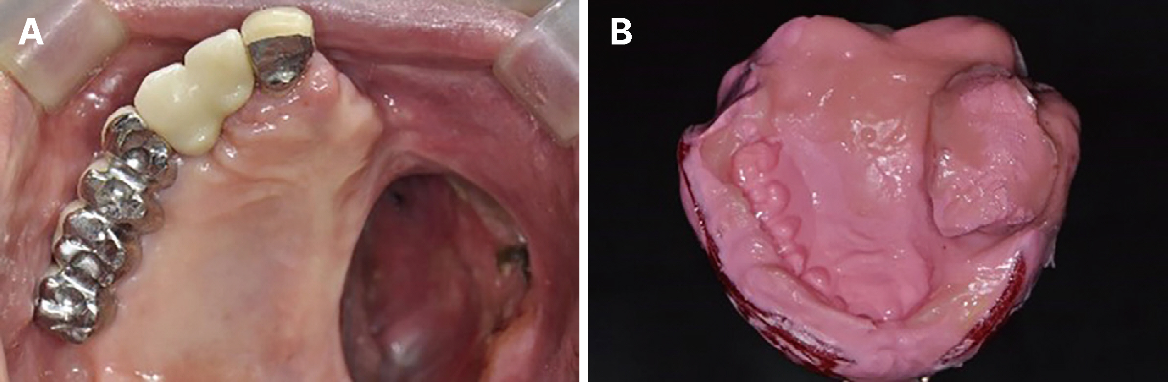
Fig. 4 Intraoral photograph and preliminary impression taking. Zirconia surveyed crown with cingulum rest was set on Rt. maxillary lateral incisor and canine. (A) Occlusal view, (B) Preliminary impression taking with alginate.

Fig. 5 Custom tray fabrication and fuctional impression taking. (A) Individual tray fabricated with 3D printer, (B) Border molding with modeling compound, (C) Functional impression taking with polysulfide rubber.
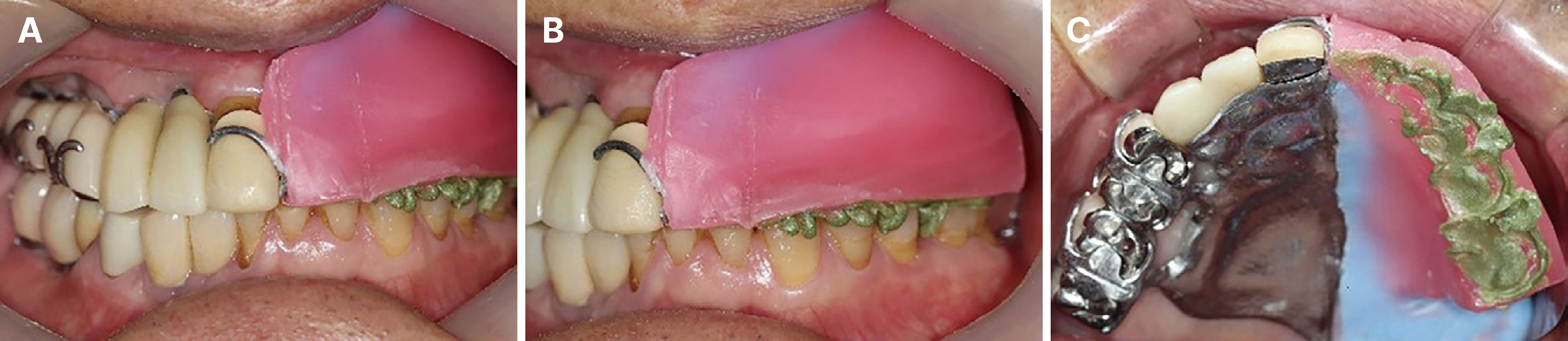
Fig. 6 Vertical dimension taking. The vertical dimension was measured based on the remaining abutment teeth. (A) Frontal view, (B) Left lateral view, (C) Occlusal view.
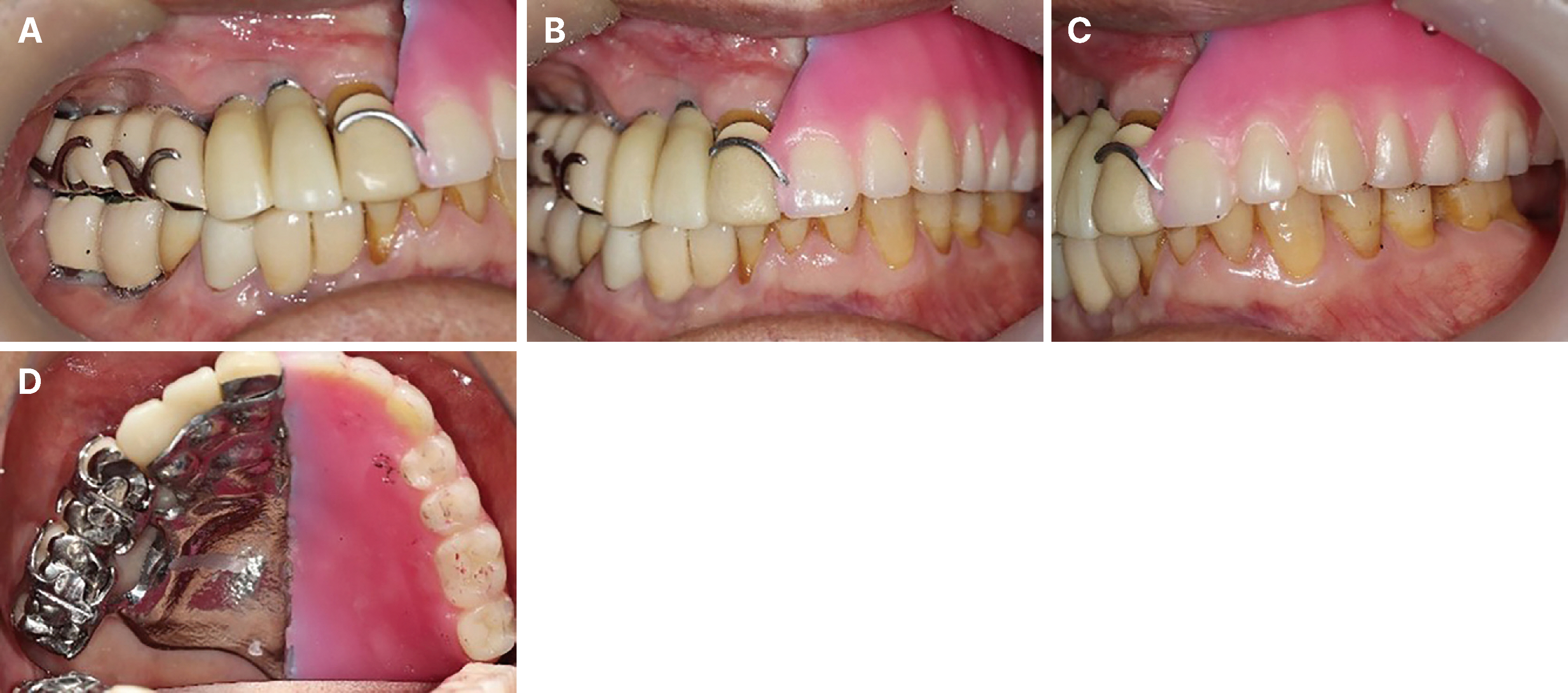
Fig. 7 Wax denture try-in. (A) Right lateral view, (B) Frontal view, (C) Left lateral view, (D) Occlusal view.
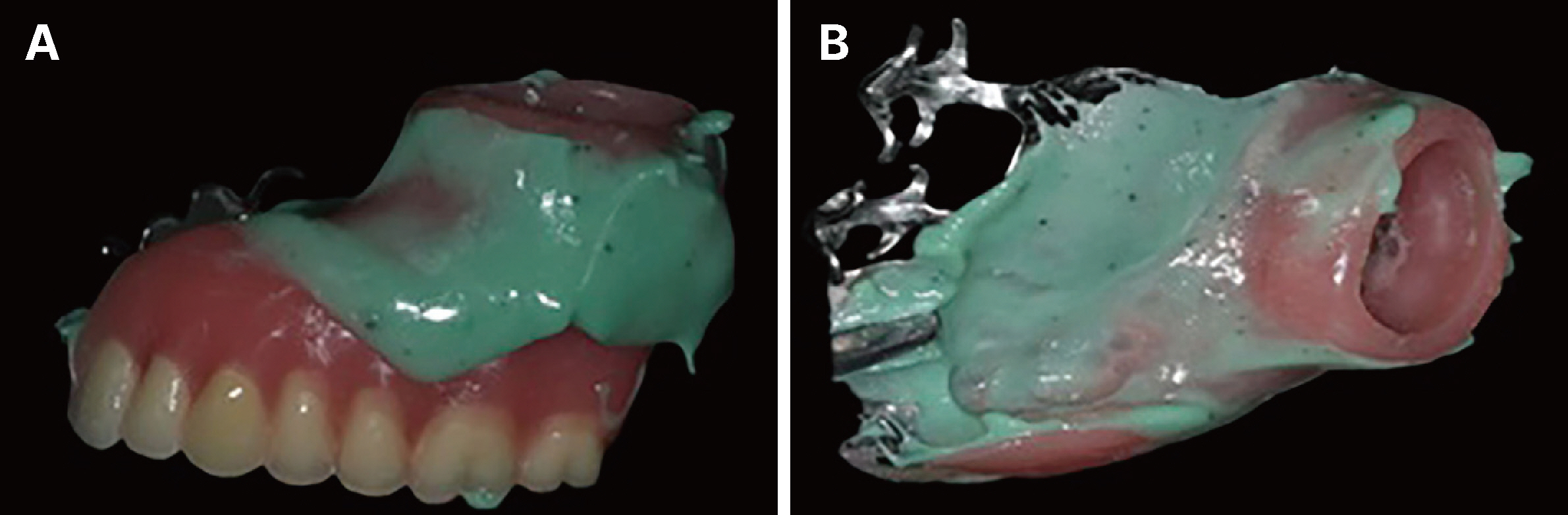
Fig. 8 Contact area checking before delivery. Pressure indicating agent was applied on the tissue surface of obturator. (A) Lateral surface, (B) Tissue surface.
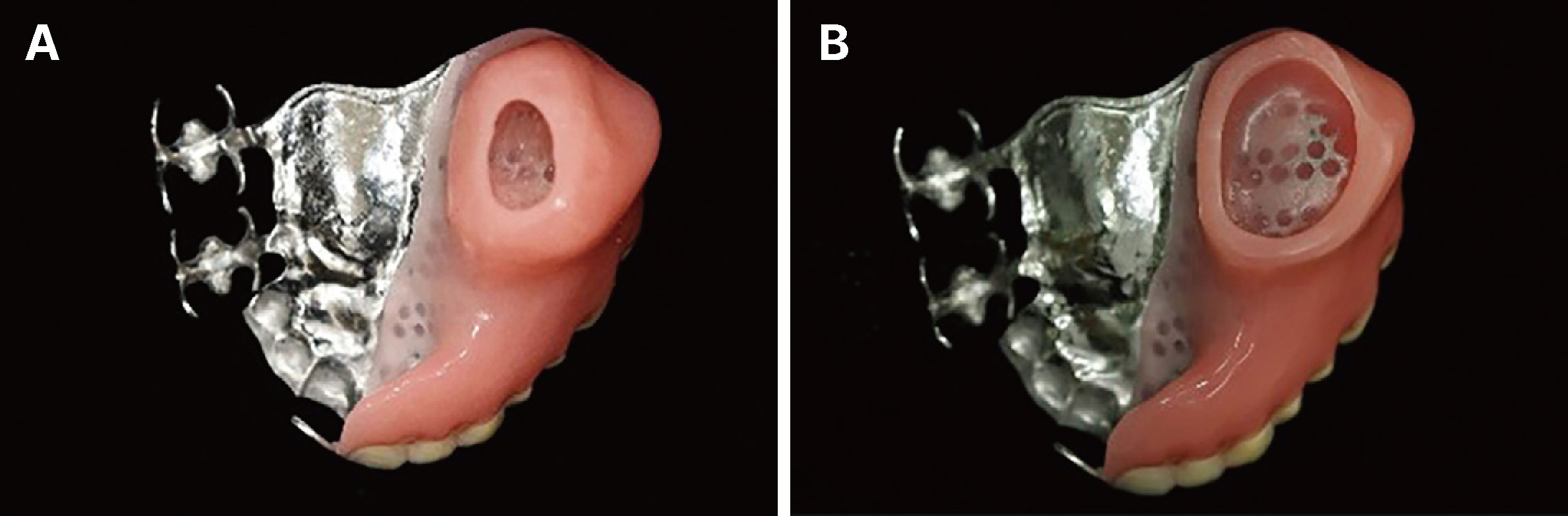
Fig. 9 Hollow bulb area checking before delivery. Before adjustment, hollow was long and heavy (A), after adjustment, hollow was much shorter and lighter than before (B). (A) Before adjustment (tissue surface), (B) After adjustment (tissue surface).
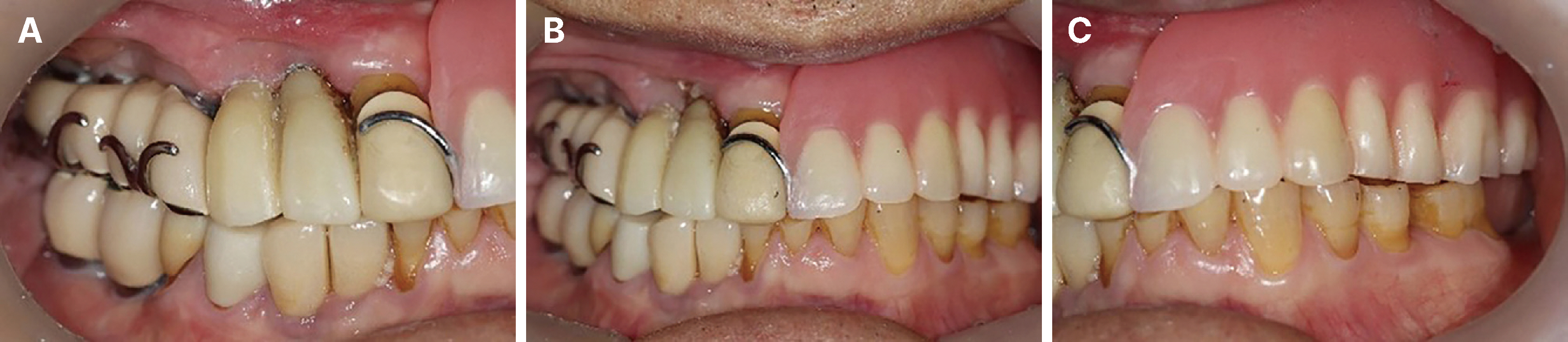
Fig. 10 Obturator was delivered and checked. (A) Right lateral view, (B) Frontal view, (C) Left lateral view.
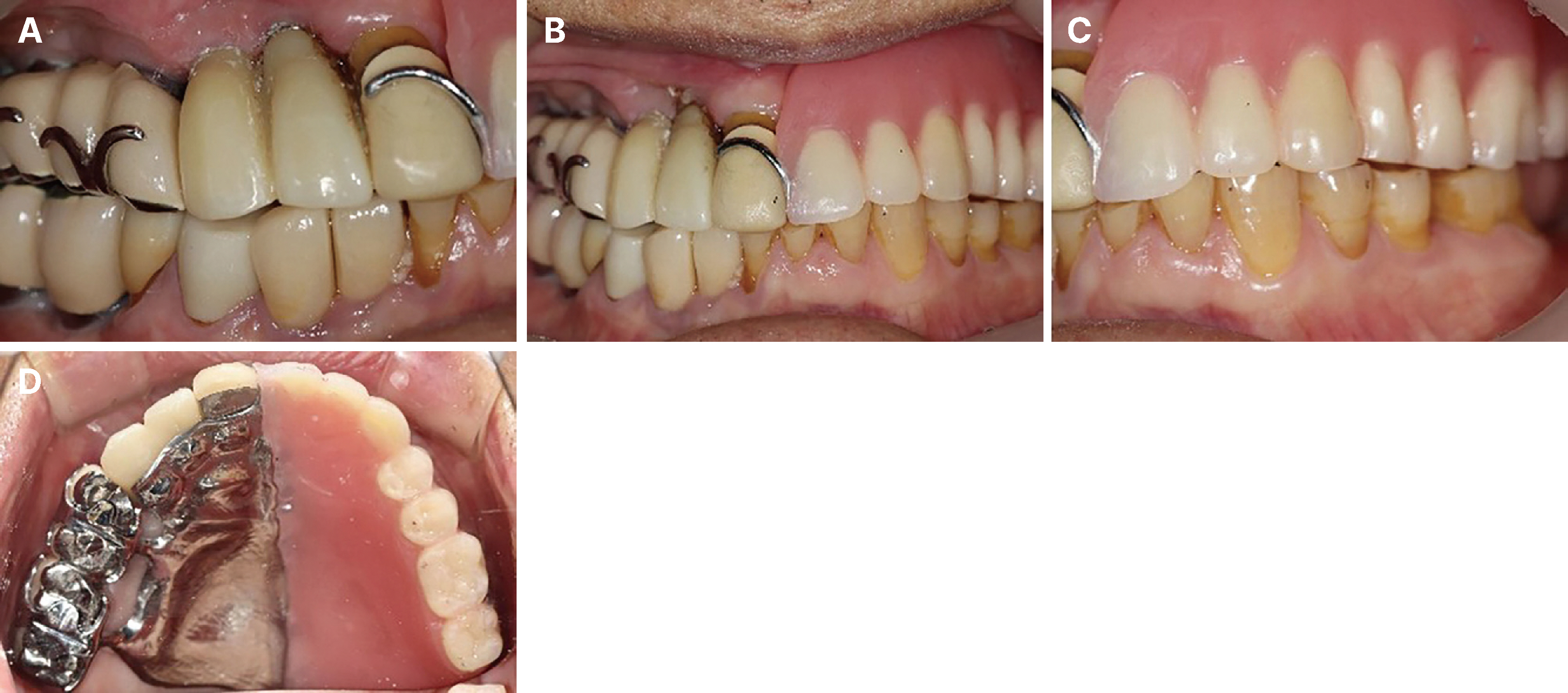
Fig. 11 Obturator was checked after 3 months. (A) Right lateral view, (B) Frontal view, (C) Left lateral view, (D) Occlusal view.
Reference
-
References
1. Tang JA, Rieger JM, Wolfaardt JF. 2008; A review of functional outcomes related to prosthetic treatment after maxillary and mandibular reconstruction in patients with head and neck cancer. Int J Prosthodont. 21:337–54. PMID: 18717093.2. Ackerman AJ. 1955; The prosthetic management of oral and facial defects following cancer surgery. J Prosthet Dent. 5:413–32. DOI: 10.1016/0022-3913(55)90050-0.3. Adell R, Lekholm U, Rockler B, Brånemark PI. 1981; A 15-year study of osseointegrated implants in the treatment of the edentulous jaw. Int J Oral Surg. 10:387–416. DOI: 10.1016/S0300-9785(81)80077-4. PMID: 6809663.4. Lyons KM, Beumer J 3rd, Caputo AA. 2005; Abutment load transfer by removable partial denture obturator frameworks in different acquired maxillary defects. J Prosthet Dent. 94:281–8. DOI: 10.1016/j.prosdent.2005.06.005. PMID: 16126081.5. Preshaw PM, Walls AWG, Jakubovics NS, Moynihan PJ, Jepson NJA, Loewy Z. 2011; Association of removable partial denture use with oral and systemic health. J Dent. 39:711–9. DOI: 10.1016/j.jdent.2011.08.018. PMID: 21924317.6. Seo H, Lee BA, Lim H, Yoon JH, Kim YT. 2019; The socioeconomic impact of Korean dental health insurance policy on the elderly: a nationwide cohort study in South Korea. J Periodontal Implant Sci. 49:248–57. DOI: 10.5051/jpis.2019.49.4.248. PMID: 31485375. PMCID: PMC6713804.7. Dilek OC, Tezulas E, Dincel M. 2007; A mini dental implant-supported obturator application in a patient with partial maxillectomy due to tumor: case report. Oral Surg Oral Med Oral Pathol Oral Radiol Endod. 103:e6–10. DOI: 10.1016/j.tripleo.2006.09.023. PMID: 17197207.8. Desjardins RP. 1978; Obturator prosthesis design for acquired maxillary defects. J Prosthet Dent. 39:424–35. DOI: 10.1016/S0022-3913(78)80161-9. PMID: 273694.9. Beumer J III, Marunick MT, Garrett N, Rohner D, Reintsema H, Abemayor E, Penn R, Nabili V, Bucher P. Beumer J, Marunick MT, Esposito SJ, editors. 2011. Rehabilitation of maxillary defects. Maxillofacial Rehabilitation: Surgical and Prosthodontic Management of Cancer-Related, Acquired, and Congenital Defects of the Head and Neck. 3rd ed. Quintessence;Chicago: p. 155–212. DOI: 10.1038/sj.bdj.2012.361.10. Aramany MA. 1978; Basic principles of obturator design for partially edentulous patients. Part I: classification. J Prosthet Dent. 40:554–7. DOI: 10.1016/0022-3913(78)90092-6. PMID: 364015.11. Pihlaja J, Näpänkangas R, Kuoppala R, Raustia A. 2015; Veneered zirconia crowns as abutment teeth for partial removable dental prostheses: a clinical 4-year retrospective study. J Prosthet Dent. 114:633–6. DOI: 10.1016/j.prosdent.2015.05.008. PMID: 26346419.
- Full Text Links
-
- Actions
-
Cited
- CITED
-
- Close
- Share
-
- Similar articles
-
- Digital interim immediate denture fabrication and implant-supported removable partial denture fabrication after multiple teeth extraction in patient with chronic periodontitis: a case report
- Repairment of abutment and abutment screw fracture in implant prosthesis: A case report
- Implant assisted obturator in patient after maxillectomy: a case report
- Immediate placement of implant following extraction of impacted supernumerary teeth and permanent teeth : A case report
- Considerations for Fabrication of CAD-CAM Abutments: Part I. Selection of Titanium Block and Fabrication Process
