Effect of surface sealant on surface roughness of dental composite with different surface roughness
- Affiliations
-
- 1Department of Conservative Dentistry, School of Dentistry, Chonnam National University, Gwangju, Republic of Korea
- 2Cheong Dental Clinic, Gwangju, Republic of Korea
- KMID: 2550428
- DOI: http://doi.org/10.14368/jdras.2023.39.4.195
Abstract
- Purpose
This study aimed to evaluate the influence of surface sealants on the surface roughness of composite resins.
Materials and Methods
The study used microfilled composite resin (Metafil CX, Sun Medical Co.) and hybrid composite resin (Aelite™ LS posterior, Bisco). Sixty specimens (8 mm in diameter and 4 mm in height) of each composite resin type were prepared and divided into 3 groups. Each specimen was ground with 600, 1000, and 2000-grit sandpaper. The Surface roughness (Ra) values were measured using a surface roughness tester (SJ-301, Mytutoyo) before and after surface sealant application. Surface sealants, BisCover™ LV (Bisco), Optiguard® (Kerr), and Seal-n-Shine™ (Pulpdent), were applied to the specimens, as instructed and observed by scanning electron microscope (JSM-7500, JEOL) and atomic force microscope (MultiMode IV, Veeco Instruments).
Results
Specimens ground with 600-grit sandpaper coated with surface sealants exhibited significantly lower Ra values than the untreated group (P < 0.05). Specimens ground with 1000 and 2000-grit sandpaper showed statistically no difference. There was no significant difference in surface roughness among BisCover™ LV, Optiguard®, and Seal-n-Shine™. SEM and AFM revealed remarkably decreased microdefects on the surfaces of composite resins after surface sealant application.
Conclusion
Surface sealants can influence surface roughness when applied on the rough surface of composite resins but not on highly polished composite resins.
Keyword
Figure
-

Fig. 1 Surface roughness (Ra, μm) of Aelite™ LS Posterior composite resins. * Statistically significant (P < 0.05).
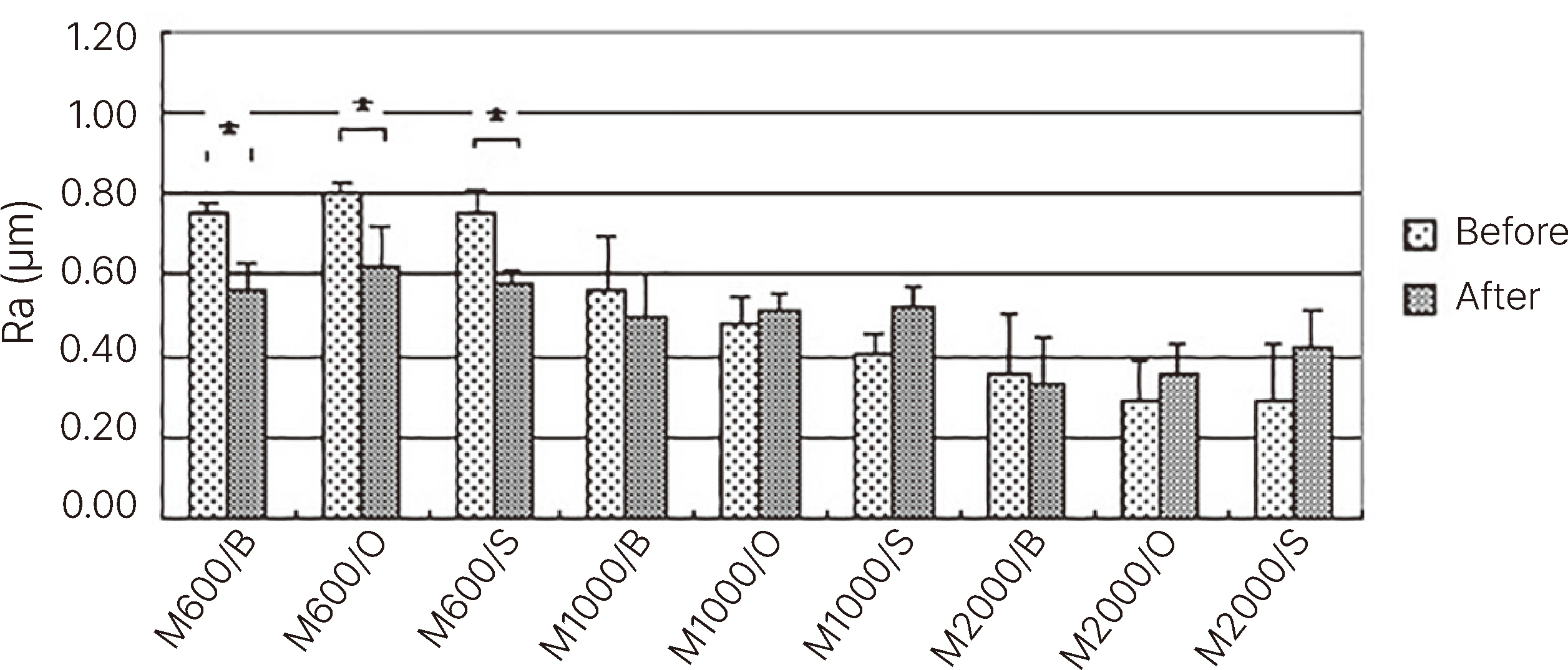
Fig. 2 Surface roughness (Ra, μm) of Metafil CX composite resins. * Statistically significant (P < 0.05).

Fig. 3 nning electron micrographs of Aelite™ LS Posterior resins that were polished with 600 grit sand paper and treated with Surface sealants (× 2500). (A) Untreated, (B) BisCover LV, (C) Optiguard, (D) Seal-n-Shine.

Fig. 4 Scanning electron micrographs of Aelite™ LS Posterior resins that were polished with 1000 grit sand paper and treated with Surface sealants (× 2500). (A) Untreated, (B) BisCover LV, (C) Optiguard, (D) Seal-n-Shine.
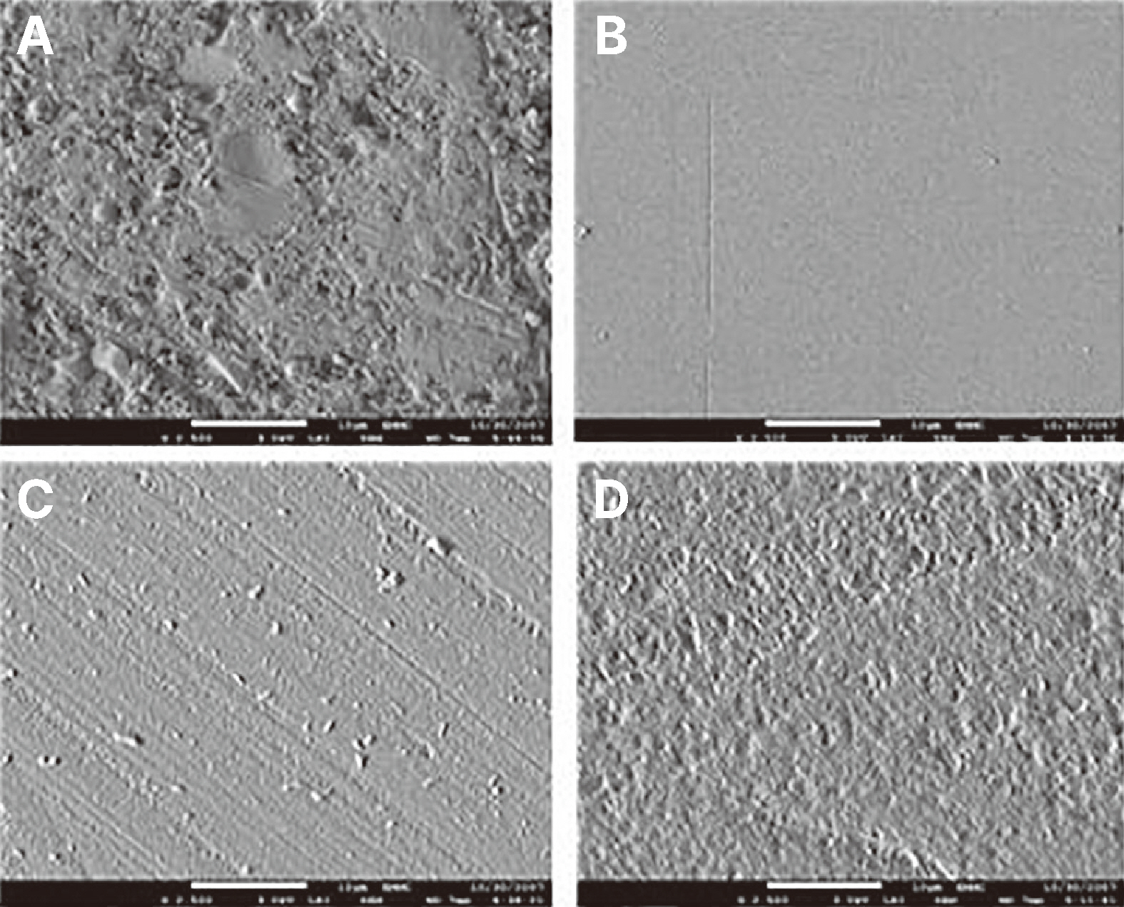
Fig. 5 Scanning electron micrographs of Aelite™ LS Posterior resins that were polished with 2000 grit sand paper and treated with Surface sealants (× 2500). (A) Untreated, (B) BisCover LV, (C) Optiguard, (D) Seal-n-Shine.
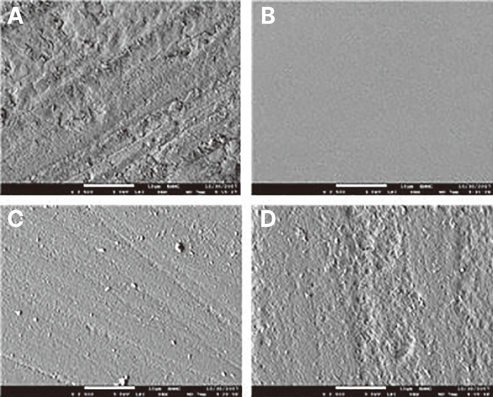
Fig. 6 Scanning electron micrographs of Metafil CX resins that were polished with 600 grit sand paper and treated with Surface sealants (× 2500). (A) Untreated, (B) BisCover LV, (C) Optiguard, (D) Seal-n-Shine.

Fig. 7 Scanning electron micrographs of Metafil CX resins that were polished with 1000 grit sand paper and treated with Surface sealants (× 2500). (A) Untreated, (B) BisCover LV, (C) Optiguard, (D) Seal-n-Shine.

Fig. 8 Scanning electron micrographs of Metafil CX resins that were polished with 2000 grit sand paper and treated with Surface sealants (× 2500). (A) Untreated, (B) BisCover LV, (C) Optiguard, (D) Seal-n-Shine.

Fig. 9 Atomic force microscopy images of Aelite™ LS Posterior resins that were polished with 2000 grit sand paper and treated with Surface sealants. (A) Untreated, (B) BisCover LV, (C) Optiguard, (D) Seal-n-Shine.
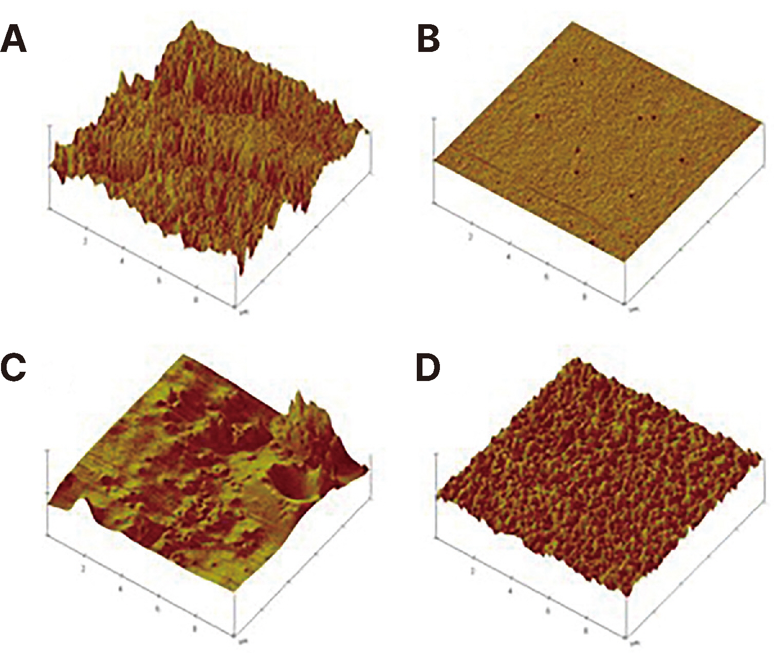
Fig. 10 Atomic force microscopy images of Metafil CX resins that were polished with 2000 grit sand paper and treated with Surface sealants. (A) Untreated, (B) BisCover LV, (C) Optiguard, (D) Seal-n-Shine.
Reference
-
References
1. Teixera EC, Thompson JL, Piascik JR, Thomson JY. 2005; In vitro toothbrush - dentifrice abrasion of two restorative composites. J Esthet Restor Dent. 17:172–80. discussion 181–2. DOI: 10.1111/j.1708-8240.2005.tb00109.x. PMID: 15996389.2. Stoddard JW, Johnson GH. 1991; An evaluation of polishing agents for composite resins. J Prosthet Dent. 65:491–5. DOI: 10.1016/0022-3913(91)90286-6. PMID: 1829763.3. Pratten DH, Johnson GH. 1988; An evaluation of finishing instruments for an anterior and a posterior composite. J Prosthet Dent. 60:154–8. DOI: 10.1016/0022-3913(88)90306-X. PMID: 2845069.4. Shinkai K, Suzuki S, Leinfelder KF, Katoh Y. 1994; Effect of surface-penetrating sealant on wear resistance of luting agents. Quintessence Int. 25:767–71. PMID: 7568681.5. Dickinson GL, Leinfelder KF, Mazer RB, Russell CM. 1990; Effect of surface penetrating sealant on wear rate of posterior composite resins. J Am Dent Assoc. 121:251–5. DOI: 10.14219/jada.archive.1990.0240. PMID: 2144862.6. Reid JS, Saunders WP, Chen YY. 1991; The effect of bonding agent and fissure sealant on microleakage of composite resin restorations. Quintessence Int. 22:295–8. PMID: 1891603.7. Ramos RP, Chinelatti MA, Chimello DT, Dibb RG. 2002; Assessing microleakage in resin composite restorations rebonded with a surface sealant and three low-viscosity resin systems. Quintessence Int. 33:450–6. PMID: 12073726.8. Sarac D, Sarac YS, Kulunk S, Ural C, Kulunk T. 2006; The effect of polishing techniques on the surface roughness and color change of composite resins. J Prosthet Dent. 96:33–40. DOI: 10.1016/j.prosdent.2006.04.012. PMID: 16872928.9. Bollen CM, Lambrechts P, Quirynen M. 1997; Comparison of surface roughness of oral hard materials to the threshold surface roughness for bacterial plaque retention: a review of the literature. Dent Mater. 13:258–69. DOI: 10.1016/S0109-5641(97)80038-3. PMID: 11696906.10. Seghi RR, Johnston WM, O'Brien WJ. 1986; Spectrophotometric analysis of color differences between porcelain systems. J Prosthet Dent. 56:35–40. DOI: 10.1016/0022-3913(86)90279-9. PMID: 3459879.11. Ramos RP, Chimello DT, Chinelatti MA, Dibb RG, Mondelli J. 2000; Effect of three surface sealants on marginal sealing of class V composite resin restorations. Oper Dent. 25:448–53. PMID: 11203855.12. Kawai K, Leinfelder KF. 1993; Effect of surface-penetrating sealant on composite wear. Dent Mater. 9:108–13. DOI: 10.1016/0109-5641(93)90085-5. PMID: 8595838.13. Rueggeberg FA, Margeson DH. 1990; The effect of oxygen inhibition on an unfilled/filled composite system. J Dent Res. 69:1652–8. DOI: 10.1177/00220345900690100501. PMID: 2212209.14. Yao K, Kohara O, Chikamori M, Kusida Y, Hieda T. 1990; SEM observation for composite resin filler. Shoni Shikagaku Zasshi. 28:918–27. PMID: 2134127.
- Full Text Links
-
- Actions
-
Cited
- CITED
-
- Close
- Share
-
- Similar articles
-
- Effect of dental bleaching on the microhardness and surface roughness of sealed composite resins
- The effects of surface glazing materials on dental sealant surface hardness and roughness
- The effect of repeated firings on the color change and surface roughness of dental ceramics
- A Study Of Surface Roughness Of Composite Resin
- Influence of the Surface roughness on translucency and surface color of the dental composite resins
