Determination of Distributional Characteristics and Efficacy of Hand Radiographic Damage in Patients with Rheumatoid Arthritis Using a Novel Damage Assessment Method
- Affiliations
-
- 1Department of Medicine, Wonkwang University School of Medicine, Iksan, Korea
- 2Division of Rheumatology, Department of Internal Medicine, Wonkwang University Hospital, Iksan, Korea
- KMID: 2549691
- DOI: http://doi.org/10.15384/kjhp.2023.23.4.209
Abstract
- Background
The purpose of this study was to identify the distribution characteristics of radiological damage to the hand of rheumatoid arthritis patients and evaluate its effectiveness using a new damage assessment method.
Methods
Hand radiographs of a total of 127 rheumatoid arthritis patients were evaluated. We simplified the pre-existing van der Heijde modified total sharp score to assess the extent of bone damage. We selected 36 joints in both hands as regions of interest in our own way, and the erosion and joint space narrowing were scored.
Results
When the erosion and joint space narrowing score values of each joint of the hand were added together, the joint space narrowing score was higher than the overall erosion score. We can see that the scaphoid,lunate,radius and ulnar joints have higher erosion scores, and the 4th proximal interphalangeal (PIP), Scaphoid, Lunate, and Radius joints have higher joint space narrowing scores than other joints. These damage assessment results were similar to the results from the previous damage assessment model.
Conclusions
The joint space narrowing score is higher than the erosion score, suggesting that joint space narrowing precedes erosion. When checking the hand X-ray of rheumatoid arthritis patients, it is necessary to carefully check the scaphoid,lunate,radius and ulnar and 4th PIP joints, which relatively high radiographic damage scores. Also, the damage assessment model used in this study is relatively simple and does not show difference in analysis results from existing assessment models, so it can be considered for application in follow-up research.
Figure
-
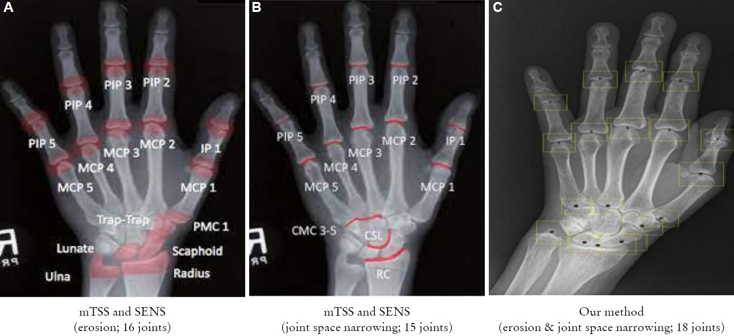
Figure 1. (A-C) Region of interest (mTSS and SENS vs. our method). PIP, proximal inter-phalageal; MCP, meta-carpo-phalangeal; IP, inter-phalangeal; PMC, proximal metacarpal; CMC, carpometacarpal; CSL, capitate-scaphoid-lunate; RC, radiocarpal; mTSS, modified total sharp score; SENS, simple erosion narrowing score.

Figure 2. (A) Total sum of each joint erosion score. (B) Total sum of each joint space narrowing score. PIP, proximal inter-phalangeal; MCP, meta-carop-phalangeal; IP, inter-phalangeal; CMC, carpo-meta-carpal; JSN_R, joint space narrowing_right; JSN_L, joint space narrowing_left.
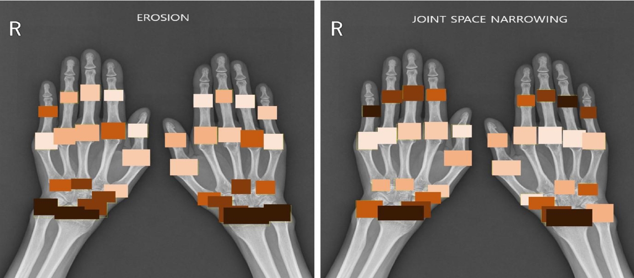
Figure 3. Heatmap of radiographic damage (darker color means more damage).
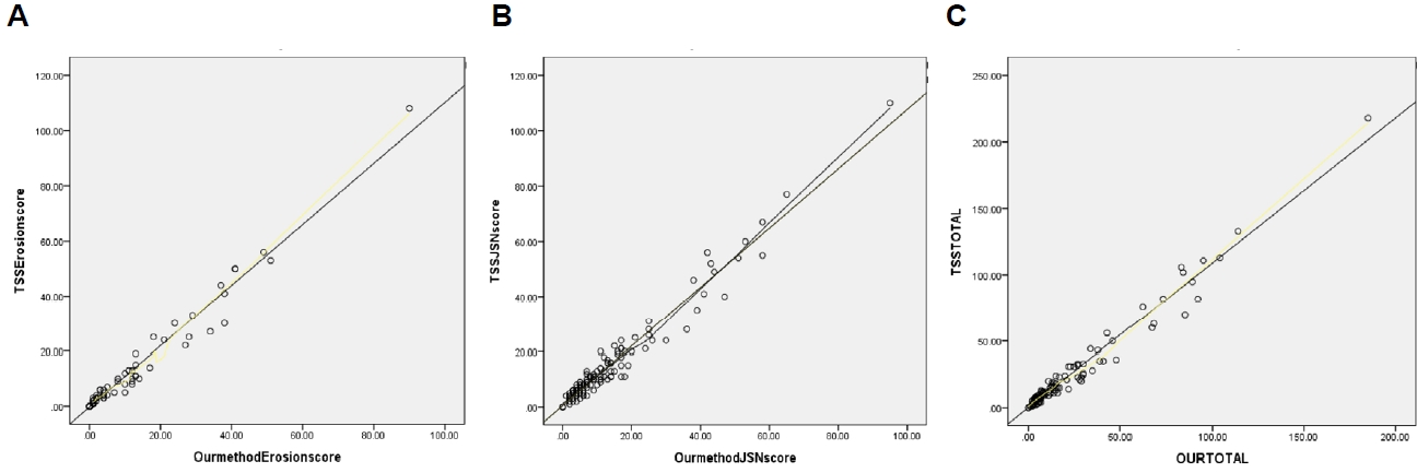
Figure 4. (A-C) Pearson’s coefficient of correlation (r) between the values of mTSS and our method score. (A) Erosion score r=0.986 (P<0.01). (B) Joint space narrowing score r=0.978 (P<0.01). (C) Total score r=0.984 (P<0.01). TSSE, total sharp score erosion; TSSJSN, total sharp score joint space narrowing; TSS, total sharp score; mTSS, modified total sharp score by van der Heijde.
Reference
-
1. Pincus T, Callahan LF. What is the natural history of rheumatoid arthritis? Rheum Dis Clin North Am. 1993; 19(1):123–51.
Article2. Scott DL, Symmons DP, Coulton BL, Popert AJ. Long-term outcome of treating rheumatoid arthritis: results after 20 years. Lancet. 1987; 1(8542):1108–11.
Article3. Wolfe F. The determination and measurement of functional disability in rheumatoid arthritis. Arthritis Res. 2002; 4(Suppl 2):11–5.4. Kanazawa T, Nishino J, Tohma S, Tanaka S. Analysis of the affected joints in rheumatoid arthritis patients in a large Japanese cohort. Mod Rheumatol. 2013; 23(1):44–9.
Article5. Plant MJ, Jones PW, Saklatvala J, Ollier WE, Dawes PT. Patterns of radiological progression in early rheumatoid arthritis: results of an 8 year prospective study. J Rheumatol. 1998; 25(3):417–26.6. Braum LS, McGonagle D, Bruns A, Philipp S, Hermann S, Aupperle K, et al. Characterisation of hand small joints arthropathy using high-resolution MRI--limited discrimination between osteoarthritis and psoriatic arthritis. Eur Radiol. 2013; 23(6):1686–93.
Article7. Colebatch AN, Edwards CJ, Østergaard M, van der Heijde D, Balint PV, D'Agostino MA, et al. EULAR recommendations for the use of imaging of the joints in the clinical management of rheumatoid arthritis. Ann Rheum Dis. 2013; 72(6):804–14.
Article8. Sudoł-Szopińska I, Matuszewska G, Pracoń G. Radiographic atlas of inflammatory rheumatic diseases. Connective tissue diseases & spondyloarthritis. 1st ed. Warsaw: Medisfera;2022. p. 21–2.9. Davies AM, Grainger AJ, James SJ. Imaging of the hand and wrist. Techniques and applications. 1st ed. Berlin: Springer;2013. p. 3–4.10. Gadeholt O, Hausotter K, Eberle H, Klink T, Pfeil A. Differing X-ray patterns in seronegative and seropositive rheumatoid arthritis. Clin Rheumatol. 2019; 38(9):2403–10.
Article11. Ory PA. Interpreting radiographic data in rheumatoid arthritis. Ann Rheum Dis. 2003; 62(7):597–604.
Article12. van der Heijde D. How to read radiographs according to the Sharp/van der Heijde method. J Rheumatol. 2000; 27(1):261–3.13. Song YH, Jun JB, Jung JH, Chang DK, Shim SC, Koh HK, et al. Radiological feature and significance of hand X-rayin early rheumatoid arthritis. Korean J Med. 1998; 55(6):1079–88.14. Salaffi F, Carotti M, Beci G, Di Carlo M, Giovagnoni A. Radiographic scoring methods in rheumatoid arthritis and psoriatic arthritis. Radiol Med. 2019; 124(11):1071–86.
Article15. Greenspan A. Erosive osteoarthritis. Semin Musculoskelet Radiol. 2013; 7(2):155–9.
Article16. Butz KD, Merrell G, Nauman EA. A biomechanical analysis of finger joint forces and stresses developed during common daily activities. Comput Methods Biomech Biomed Engin. 2012; 15(2):131–40.
Article17. Koh JH, Jung SM, Lee JJ, Kang KY, Kwok SK, Park SH, et al. Radiographic structural damage is worse in the dominant than the non-dominant hand in individuals with early rheumatoid arthritis. PLoS One. 2015; 10(8):e0135409.
Article
- Full Text Links
-
- Actions
-
Cited
- CITED
-
- Close
- Share
-
- Similar articles
-
- Predictors of joint damage in patients with rheumatoid arthritis: focus on short- and long-term effects of intra-articular glucocorticoid injections
- Correlation of Anti-Cyclic Citrullinated Antibody with Hand Joint Erosion Score in Rheumatoid Arthritis Patients
- Rheumatoid Hand Surgery in the Era of Biologic Therapy: A Rheumatologist-oriented Overview
- S100A8/A9 as a biomarker for synovial inflammation and joint damage in patients with rheumatoid arthritis
- Bone Loss and Radiographic Damage Profile in Rheumatoid Arthritis Moroccan Patients
