Using treatment denture and digital technology in patient with unstable mandibular movement: a case report
- Affiliations
-
- 1Department of Prosthodontics, School of Dentistry, Chonnam National University, Gwangju, Republic of Korea
- KMID: 2549283
- DOI: http://doi.org/10.14368/jdras.2023.39.3.168
Abstract
- Long-term use of inappropriate prosthesis often results in habitual closure or irregular mandibular movements. In that case, guide to the stable centric position is difficult. Therefore, by using a treatment denture, the muscles and TMJ should be stabilized and the jaw relation should be acquired with the treatment position. Compared to the conventional method, digital technology in fabrica-tion complete denture has the advantage of reproducing a stable tooth arrangement in cases of difficult tooth alignment, minimiz-ing laboratory errors and reducing denture fabrication time. Therefore, in this case, the final denture was fabricated by digitally re-producing the stable treatment position, vertical dimension, and lip support with a treatment denture, and satisfactory results were obtained.
Figure
-

Fig. 1 Initial radiograph. (A) Panoramic radiograph, (B) TMJ radiograph. flattened condyle head.
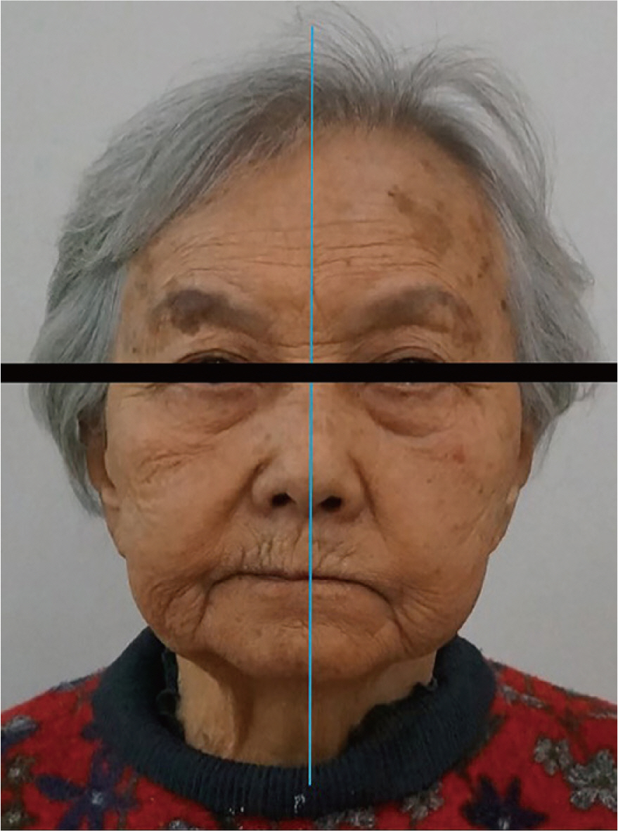
Fig. 2 Extraoral photograph. It shows mandibular deviation to right side.
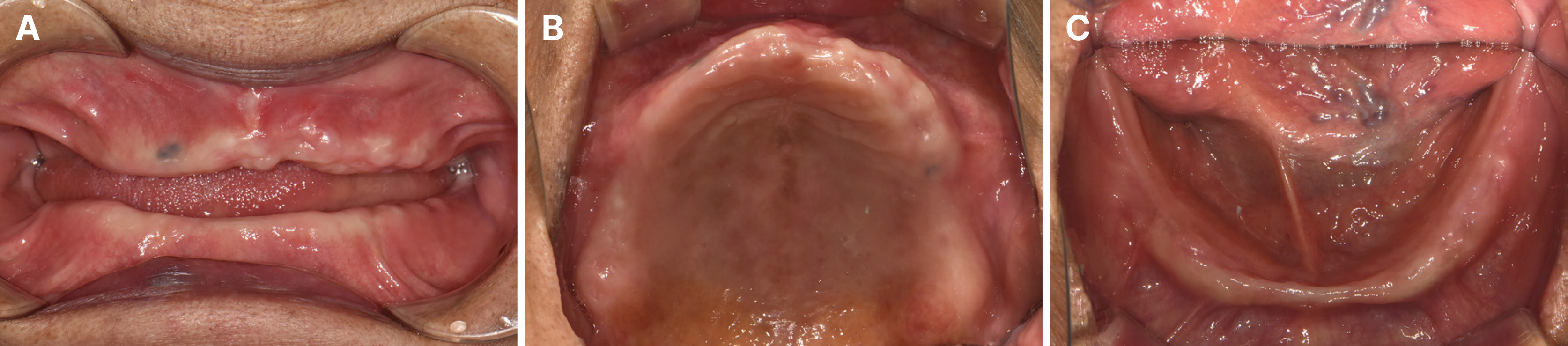
Fig. 3 Intraoral photographs (Post-extraction). (A) Maxillary occlusal view, (B) Frontal view, (C) Mandibular occlusal view.

Fig. 4 VD and CR registration. (A) Gothic arch tracing, (B) Mounted in the articulator.

Fig. 5 Fabrication of treatment denture. (A) Mixed talcum powder and self cure resin, (B) Flat occlusal table treatment denture.

Fig. 6 Indentation marks on the flat occlusal table of treatment denture (A) First time delivery. It shows spread mark, (B) 4 weeks after delivery. It shows repeatable mark.

Fig. 7 A series of Processes to obtain definitive digital cast. (A) Functional impressions taken with the treatment dentures, (B) Inverting of denture impression phase using CAD software(Meshmixer, Autodesk Inc, San Jose, USA), (C) Determination of jaw relation with treatment denture, (D) Definitive digital cast.
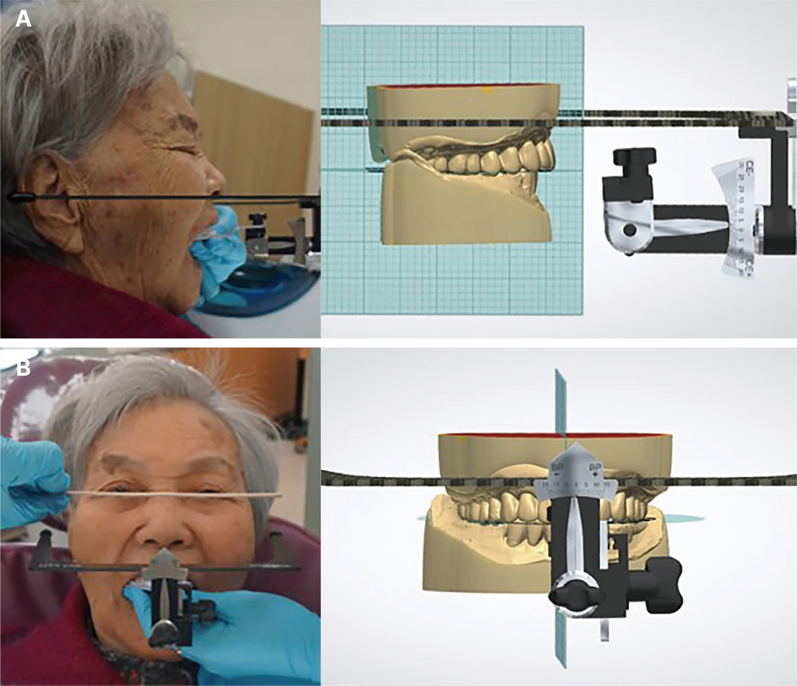
Fig. 8 Digital facebow transfer with UTS CAD. (A) Parallel to the camper’s plane (CE: -3.0), (B) Parallel to the interpupillary line (BP: -1.0).

Fig. 9 Digital mounting on the virtual articulator. (A) Determination of occlusal plane with treatment denture, (B) Teeth arrangement on the tissue surface, (C) Facial scan data as a reference of teeth arrangement.

Fig. 10 3D printed trial denture. (A) Arrangement with anatomical teeth, (B) Arrangement with nonanatomical teeth, (C) Occlusal surface of anatomical teeth, (D) Occlusal surface of nonanatomical teeth.
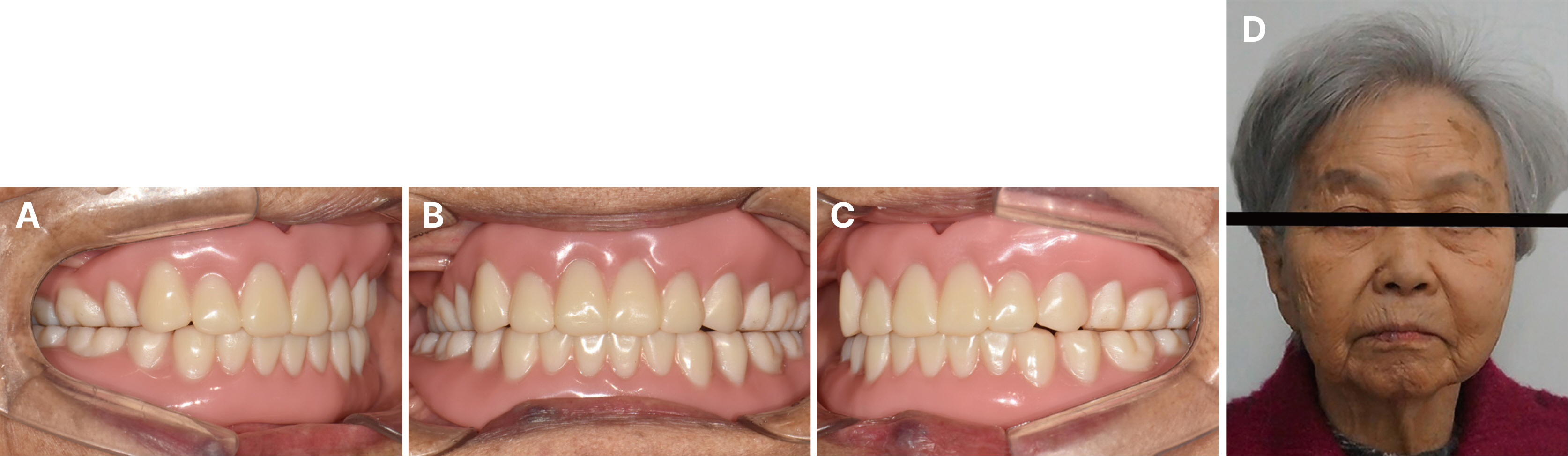
Fig. 11 Definitive denture. (A) Right lateral view, (B) Frontal view, (C) Left lateral view, (D) Facial appearance after delivery.
Reference
-
References
1. Watanabe Y. 2004; Observation of horizontal mandibular positions in an edentulous patient using a digital gothic arch tracer: a clinical report. J Prosthet Dent. 91:15–9. DOI: 10.1016/j.prosdent.2003.10.009. PMID: 14739888.2. Bidra AS, Taylor TD, Agar JR. 2013; Computer-aided technology for fabricating complete dentures: systematic review of historical background, current status, and future perspectives. J Prosthet Dent. 109:361–6. DOI: 10.1016/S0022-3913(13)60318-2. PMID: 23763779.3. Miyazaki T, Hotta Y, Kunii J, Kuriyama S, Tamaki Y. 2009; A review of dental CAD/CAM: current status and future perspectives from 20 years of experience. Dent Mater J. 28:44–56. DOI: 10.4012/dmj.28.44. PMID: 19280967.4. Slade GD. 1997; Derivation and validation of a short-form oral health impact profile. Community Dent Oral Epidemiol. 25:284–90. DOI: 10.1111/j.1600-0528.1997.tb00941.x. PMID: 9332805.5. Yoshidome K, Torii M, Kawamura N, Shimpo H, Ohkubo C. 2021; Trueness and fitting accuracy of maxillary 3D printed complete dentures. J Prosthodont Res. 65:559–64. DOI: 10.2186/jpr.JPR_D_20_00240. PMID: 33980786.6. Mercado MD, Faulkner KD. 1991; The prevalence of craniomandibular disorders in completely edentulous denture-wearing subjects. J Oral Rehabil. 18:231–42. DOI: 10.1111/j.1365-2842.1991.tb00052.x. PMID: 1856775.7. Gilboe DB. 1983; Centric relation as the treatment position. J Prosthet Dent. 50:685–9. DOI: 10.1016/0022-3913(83)90211-1. PMID: 6580438.8. Inada M, Yamazaki T, Shinozuka O, Sekiguchi G, Tamamori Y, Ohyama T. 2002; Complete denture treatments for a cerebral palsy patient by using a treatment denture. A case report. J Med Dent Sci. 49:171–7.9. Schweiger J, Güth JF, Edelhoff D, Stumbaum J. 2017; Virtual evaluation for CAD-CAM-fabricated complete dentures. J Prosthet Dent. 117:28–33. DOI: 10.1016/j.prosdent.2016.05.015. PMID: 27492983.10. Jones PM. 1972; The monoplane occlusion for complete dentures. J Am Dent Assoc. 85:94–100. DOI: 10.14219/jada.archive.1972.0293. PMID: 4503599.11. Yoshidome K, Torii M, Kawamura N, Shimpo H, Ohkubo C. 2021; Trueness and fitting accuracy of maxillary 3D printed complete dentures. J Prosthodont Res. 65:559–64. DOI: 10.2186/jpr.JPR_D_20_00240. PMID: 33980786.
- Full Text Links
-
- Actions
-
Cited
- CITED
-
- Close
- Share
-
- Similar articles
-
- Utilization of digital technology in fabricating mandibular implant overdenture for skeletal class II edentulous patient: A case report
- Full mouth rehabilitation in edentulous patient with unstable mandibular position using flat table treatment dentures and CAD-CAM technology
- Digital interim immediate denture fabrication and implant-supported removable partial denture fabrication after multiple teeth extraction in patient with chronic periodontitis: a case report
- Posterior rehabilitation considering mandibular movement with digital facebow transfer and virtual articulator: A case report
- Full mouth rehabilitation in partially edentulous patient with an unstable mandibular position
