Esthetic restoration of maxillary anterior fixed prosthesis using a digital diagnostic wax-up: a case report
- Affiliations
-
- 1Department of Prosthodontics, School of Dentistry, Chonnam National University, Gwangju, Republic of Korea
- KMID: 2549276
- DOI: http://doi.org/10.14368/jdras.2023.39.2.89
Abstract
- Recently, computer-aided design and computer-aided manufacturing (CAD/CAM) environment have changed the clinician treatment method in the fabrication of prosthesis. The diagnostic wax-up by using digital technology simplifies the laboratory process and provides clinical efficiency and convenience. In this case, Digital diagnostic wax-up was superimposed on extra-oral photo to consider the patient’s facial appearance and utilized to produce the final prosthesis. It can be more efficient and esthetic than a diagnostic wax-up that was made only on a model. The digital diagnostic wax-up that superimposed on extra-oral photo not only visualizes the predicted outcome of prosthodontic treatment but also improves satisfaction and facilitates the communication between patient and dentist. We report aesthetically and functionally satisfactory results that obtained after restoration.
Keyword
Figure
-
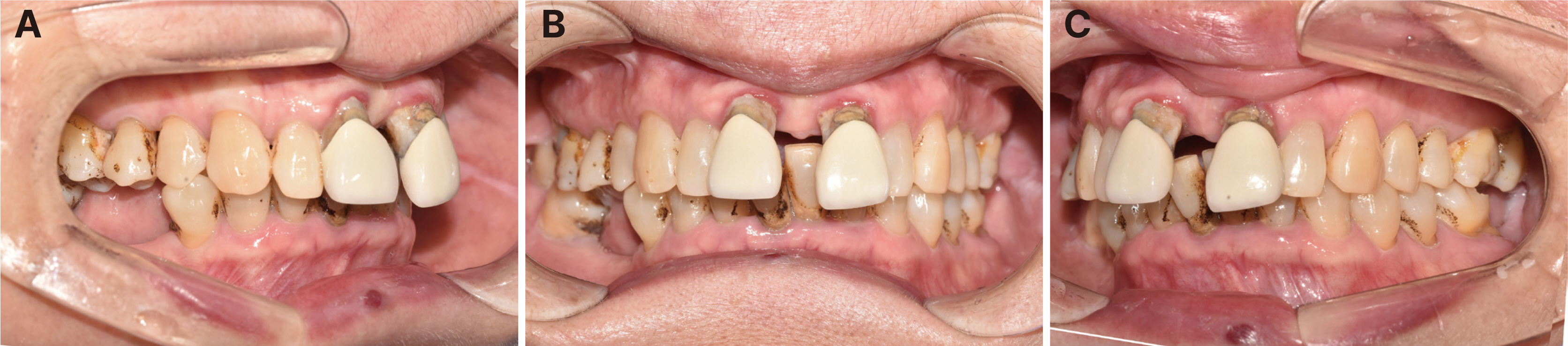
Fig. 1 Initial intraoral photographs. (A) Right lateral view, (B) Frontal view, (C) Left lateral view.
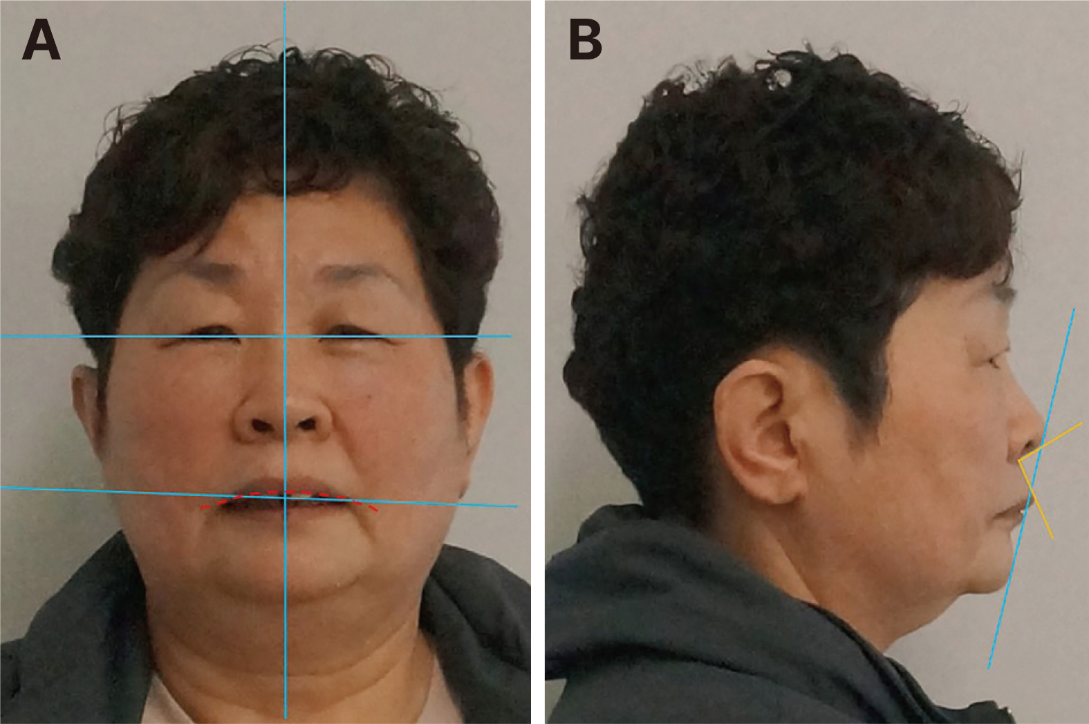
Fig. 2 Extra-oral photographs for esthetic analysis. (A) Frontal view. Right slanted incisal plane, discrepancy between facial midline and dental midline, low smile line, (B) Lateral view. Upper lip to E-line (+2 mm), 99° nasolabial angle.
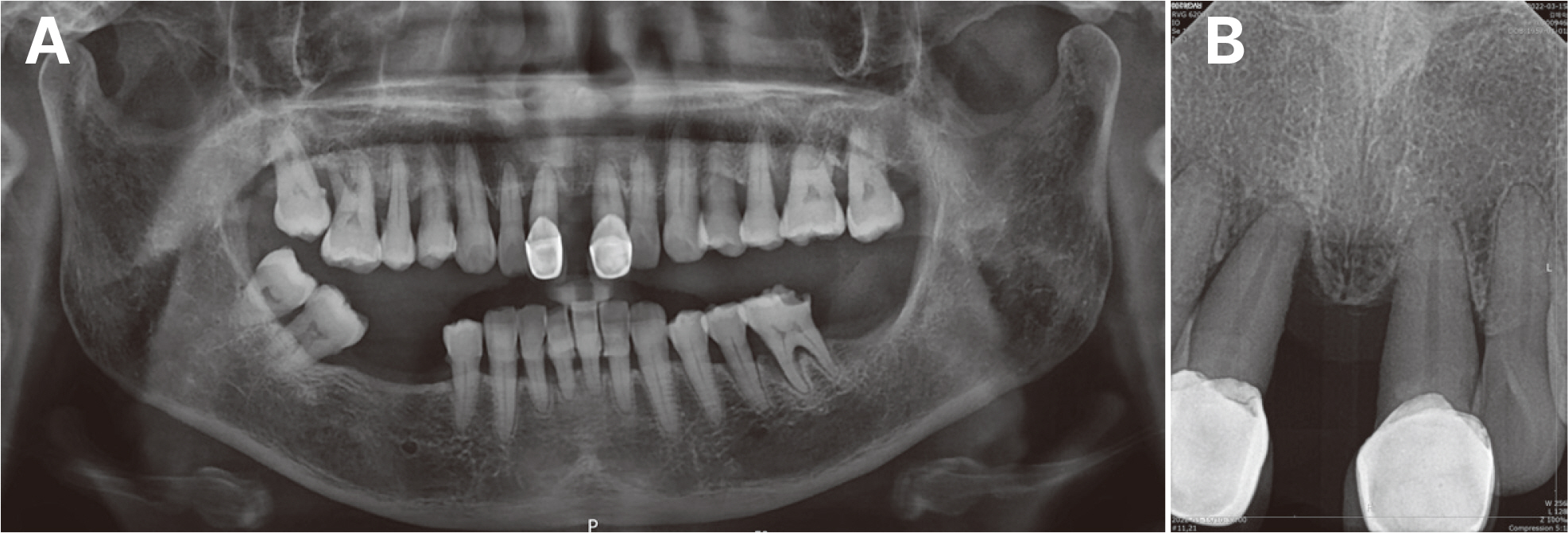
Fig. 3 Initial radiograph. (A) Panoramic radiograph, (B) Standard radiograph.
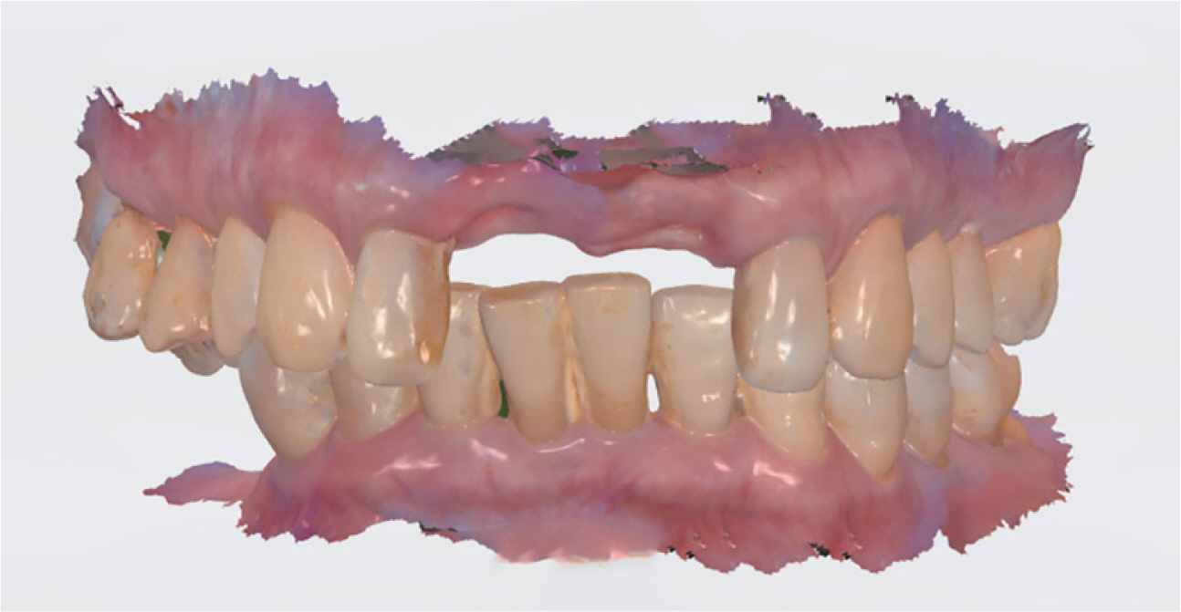
Fig. 4 Intraoral scan after extraction.
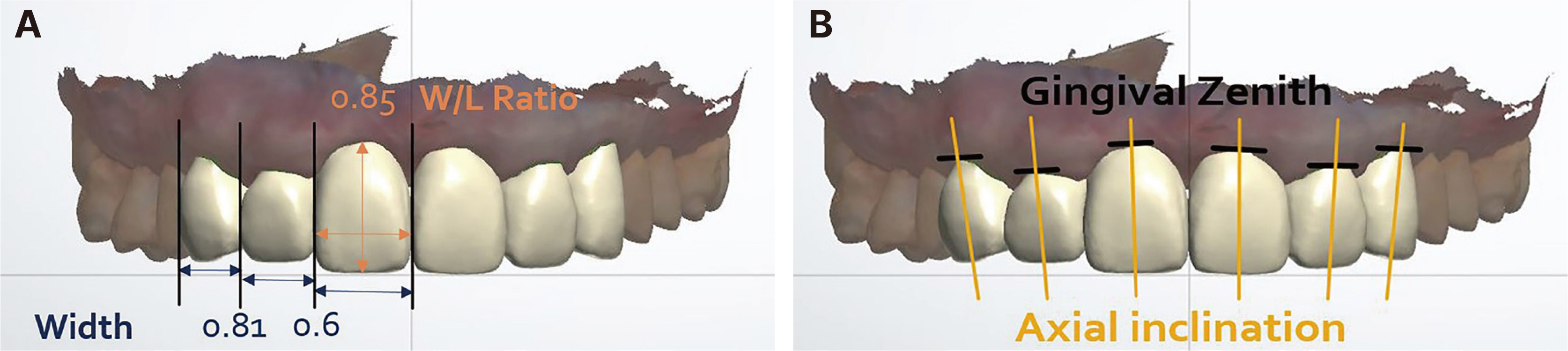
Fig. 5 Digital diagnostic wax-up on CAD software. (A) Design in consideration of tooth ratio, (B) Tooth alignment.
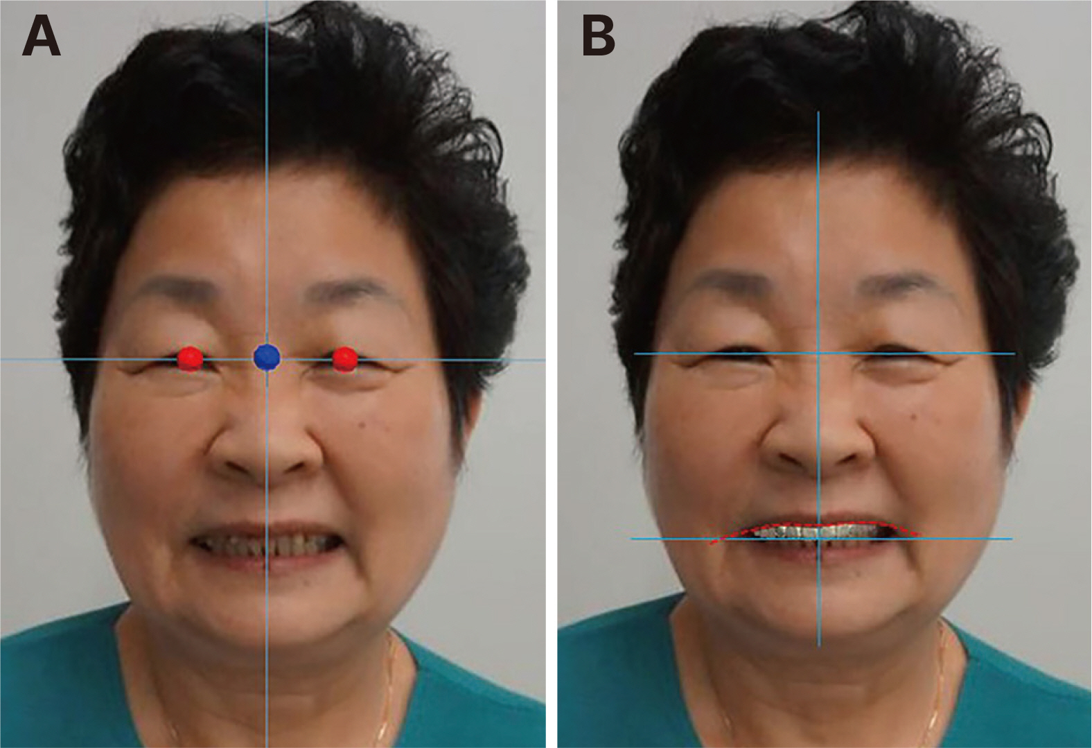
Fig. 6 Superimposition of extra-oral photographs. (A) Superimposition of digital diagnostic wax up and extra-oral photographs, (B) Esthetic re-evaluation of diagnostix wax-up.
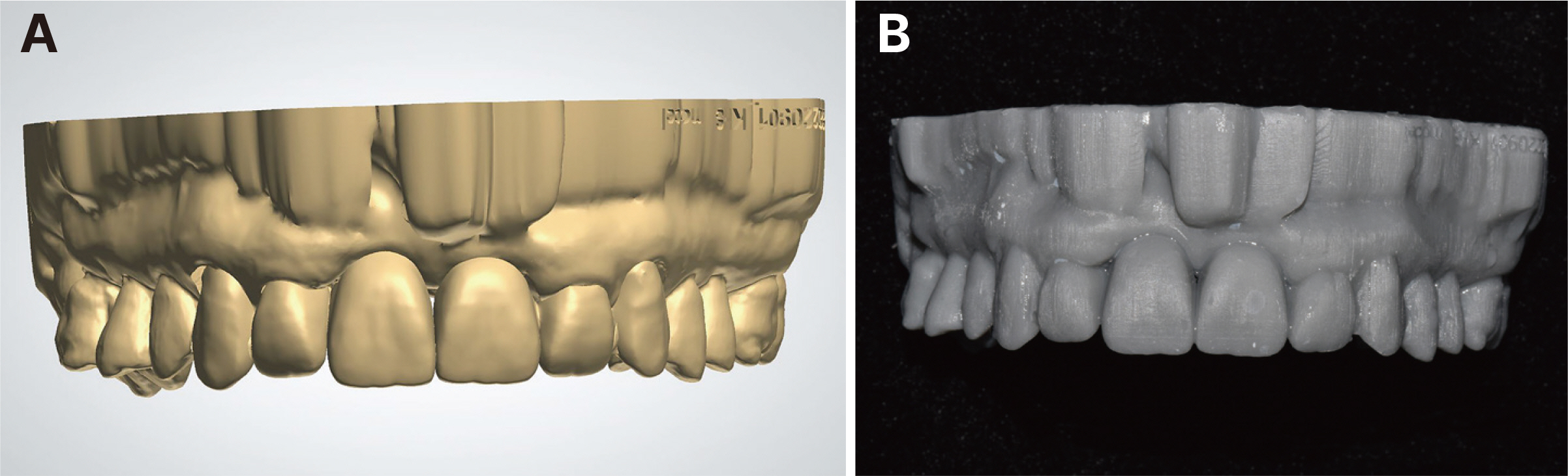
Fig. 7 Materials for counseling. (A) Digital diagnostix wax-up model, (B) 3D printed model.
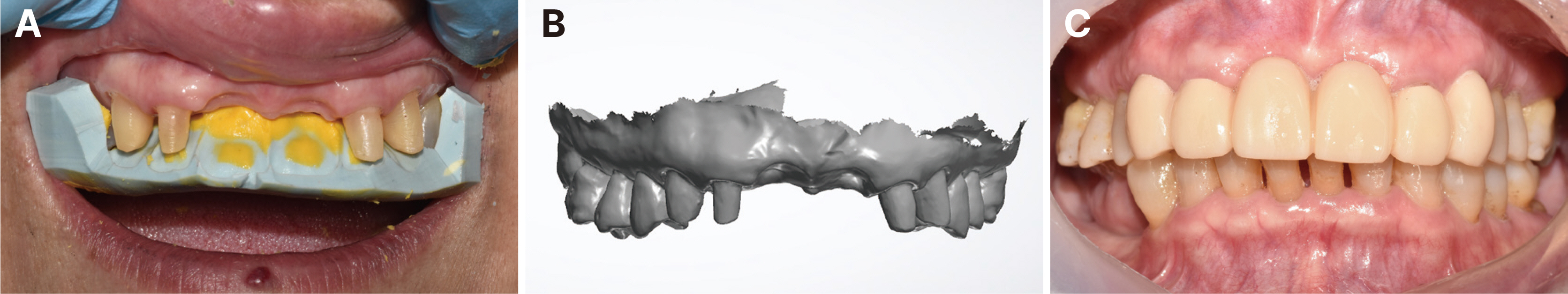
Fig. 8 Preparation. (A) Putty index for reduction guide, (B) Impression taking with intraoral scan, (C) Intraoral photo of pre-fabricated provisional restoration.
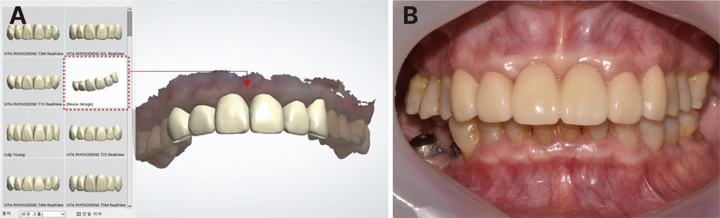
Fig. 9 Definitive prosthesis. (A) Diagnostic wax-up library, (B) Intraoral photo of final restoration.
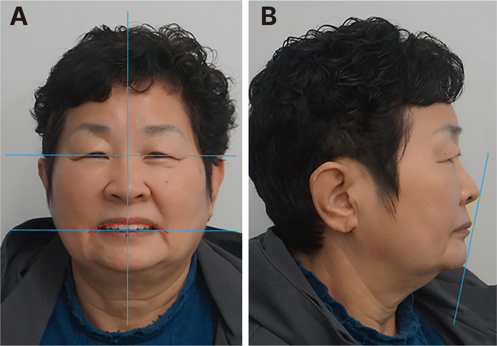
Fig. 10 Extra-oral photo of patient with final restoration. (A) Frontal view. Incisal plane parallel to inter-pupillary line, dental midline correspond to facial midline, average smile line, (B) Lateral view. Upper lip to E-line (-1 mm), 105° nasolabial angle
Reference
-
References
1. 2017; The glossary of prosthodontic terms: ninth edition. J Prosthet Dent. 117:e1–e105. DOI: 10.1016/j.prosdent.2016.12.001. PMID: 28418832.2. Morgan DW, Comella MC, Staffanou RS. 1975; A diagnostic wax-up technique. J Prosthet Dent. 33:169–77. DOI: 10.1016/S0022-3913(75)80107-7. PMID: 1090726.
Article3. Abduo J. 2016; Morphological Symmetry of Maxillary Anterior Teeth before and after Prosthodontic Planning: Comparison between Conventional and Digital Diagnostic Wax-Ups. Med Princ Pract. 25:276–81. DOI: 10.1159/000444323. PMID: 26828666. PMCID: PMC5588395.
Article4. Drafta S, Perieanu VS, Costea R, Eftene O, Burlibasa M, Manea RM, Caministeanu F, Carabela M, Neagoe IC, Costea R, Beuran IA, Maru N. 2022; Diagnostic Wax-Up - an extremely important method of communication between the dental office and the dental laboratory. Roman J Med Pract. 17:66–71. DOI: 10.37897/RJMP.2022.2.2. PMID: d74d75af915343cdba38ee6a8092a5f8.
Article5. Stern N, Brayer L. 1975; Collapse of the occlusion - aetiology, symptomatology and treatment. J Oral Rehabil. 2:1–19. DOI: 10.1111/j.1365-2842.1975.tb00907.x. PMID: 1056439.
Article6. Brunsvold MA. 2005; Pathologic tooth migration. J Periodontol. 76:859–66. DOI: 10.1902/jop.2005.76.6.859. PMID: 15948679.
Article7. Oh YA, Yang HS, Park SW, Lim HP, Yun KD, Park C. 2017; Analysis of the width ratio and wear rate of maxillary anterior teeth in the Korean population. J Adv Prosthodont. 9:93–8. DOI: 10.4047/jap.2017.9.2.93. PMID: 28435617. PMCID: PMC5397594.
Article8. Levine JB. 1995; Esthetic diagnosis. Curr Opin Cosmet Dent. 9–17. PMID: 7550888.9. Sghaireen MG, Al-Omiri MK. 2016; Relationship between impact of maxillary anterior fixed prosthodontic rehabilitation on daily living, satisfaction, and personality profiles. J Prosthet Dent. 115:170–6. DOI: 10.1016/j.prosdent.2015.07.009. PMID: 26443068.
Article10. Bidra AS. 2011; Three-Dimensional Esthetic Analysis in Treatment Planning for Implant-Supported Fixed Prosthesis in the Edentulous Maxilla: Review of the Esthetics Literature. J Esthet Restor Dent. 23:219–36. DOI: 10.1111/j.1708-8240.2011.00428.x. PMID: 21806753.
Article
- Full Text Links
-
- Actions
-
Cited
- CITED
-
- Close
- Share
-
- Similar articles
-
- Esthetic restoration of maxillary anterior teeth considering facial features in digital diagnostic wax-up: a case report
- Usage of digital technique to facilitate communication between dentist, dental lab technician, and patients in diagnosis and restoration for maxillary anterior implant: a case report
- Using dental virtual patients with dynamic occlusion in esthetic restoration of anterior teeth: case reports
- Decoronation and implant restoration of ankylosed tooth resulted from anterior avulsion: A case report
- Use of Digital Smile Design in esthetic restoration in anterior teeth: A case report
