Korean J Sports Med.
2023 Dec;41(4):201-206. 10.5763/kjsm.2023.41.4.201.
Can Suprascapular Venous Engorgement with a Paralabral Cyst in the Shoulder Aid the Diagnosis of Suprascapular Neuropathy?: A Cohort Study of Level of Evidence III
- Affiliations
-
- 1Department of Orthopedic Surgery, The Jain Hospital, Goyang, Korea
- 2Center for Shoulder, Elbow and Sports, Neon Orthopedic Clinic, Seoul, Korea
- KMID: 2548993
- DOI: http://doi.org/10.5763/kjsm.2023.41.4.201
Abstract
- Purpose
Suprascapular neuropathy can be caused by a solid mass, transverse scapular ligament hypertrophy, paralabral cyst, or dilatation of a suprascapular vein. Studies have measured the size of the cyst and veins using magnetic resonance imaging (MRI) to aid diagnosis of suprascapular neuropathy. However, it is often difficult to determine the size of a cyst and vein. we measured cyst and vein size as potential diagnostic markers for suprascapular neuropathy.
Methods
This study retrospectively enrolled 118 patients diagnosed with a paralabral cyst in a clinic from January 2016 to December 2019. After excluding other neuropathies and cysts not related to the course of the suprascapular nerve, a total of 67 patients were analyzed. The cyst diameter, cyst volume, and vein diameter were measured engorgement by MRI in axial, coronal, and sagittal T2-weighted images. Cutoff values were established based on Youden’s index.
Results
There was no significant difference between the neuropathy and control groups in cyst coronal diameter, but the neuropathy group had greater sagittal (p=0.001), axial (p=0.001), and maximum cyst diameters (p=0.005), cyst volume (p=0.003), and coronal (p=0.002), axial (p=0.001), and maximum vein diameters (p=0.001).
Conclusion
In suprascapular neuropathy symptomatic patients, electromyography/nerve conduction velocity tests are eventually needed when in doubt. However, measuring cyst diameter, volume, and suprascapular vein diameter as a screening test could be considered.
Figure
-
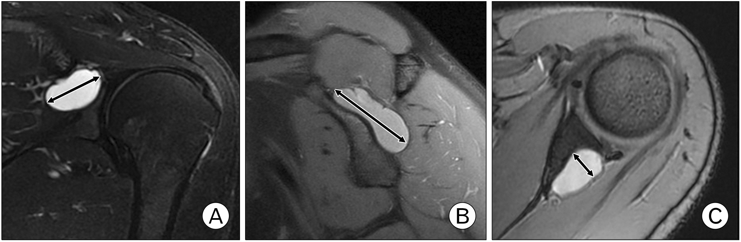
Fig. 1 Diameter of a paralabral cyst (bidirectional arrows) on magnetic resonance images in the left shoulder of a 23-year-old man. (A) Coronal oblique view, 17.1 mm. (B) Sagittal oblique view, 24.4 mm. (C) Axial view, 8.7 mm.
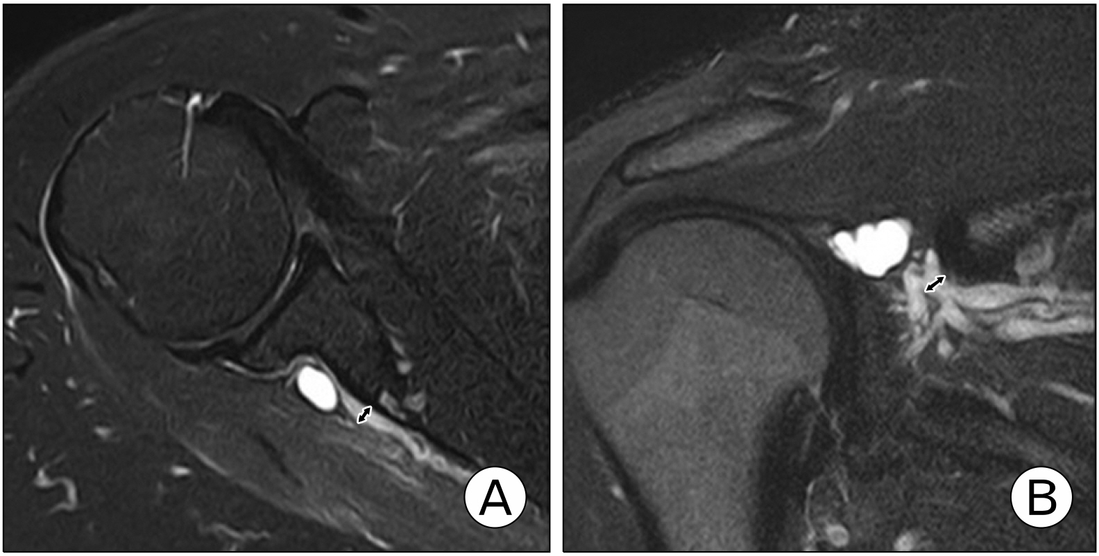
Fig. 2 Diameter of suprascapular veins (bidirectional arrows) on magnetic resonance images in the right shoulder of a 16-year-old man. (A) Axial view, 3.8 mm. (B) Coronal oblique view, 4.5 mm.
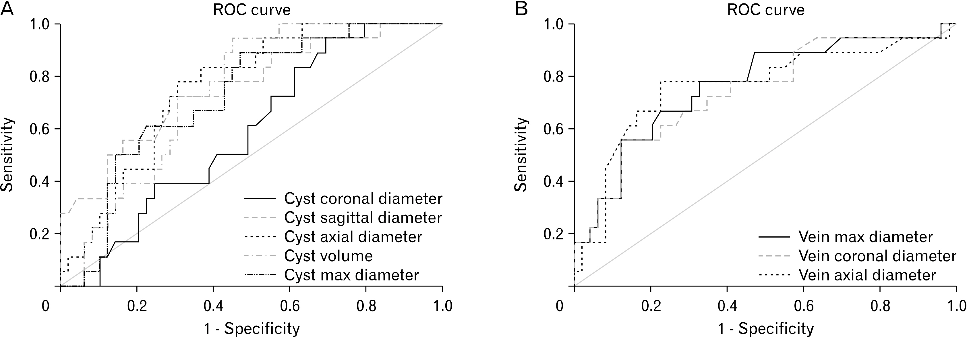
Fig. 3 (A) Receiver operating characteristic (ROC) curves of cyst coronal diameter, cyst sagittal diameter, cyst axial diameter, cyst volume, and cyst max diameter. (B) ROC curves of suprascapular vein max diameter, suprascapular vein coronal diameter, and suprascapular vein axial diameter.
Reference
-
1. Bozzi F, Alabau-Rodriguez S, Barrera-Ochoa S, et al. 2020; Suprascapular neuropathy around the shoulder: a current concept review. J Clin Med. 9:2331. DOI: 10.3390/jcm9082331. PMID: 32707860. PMCID: PMC7465639.
Article2. Kostretzis L, Theodoroudis I, Boutsiadis A, Papadakis N, Papadopoulos P. 2017; Suprascapular nerve pathology: a review of the literature. Open Orthop J. 11:140–53. DOI: 10.2174/1874325001711010140. PMID: 28400882. PMCID: PMC5366386.
Article3. Łabętowicz P, Synder M, Wojciechowski M, et al. 2017; Protective and predisposing morphological factors in suprascapular nerve entrapment syndrome: a fundamental review based on recent observations. Biomed Res Int. 2017:4659761. DOI: 10.1155/2017/4659761. PMID: 28691025. PMCID: PMC5485264.
Article4. Tung GA, Entzian D, Stern JB, Green A. 2000; MR imaging and MR arthrography of paraglenoid labral cysts. AJR Am J Roentgenol. 174:1707–15. DOI: 10.2214/ajr.174.6.1741707. PMID: 10845511.
Article5. Guven F, Ogul H, Kaya S, Kantarci M. 2020; MR arthrographic evaluation of posterior paraglenoid labral cysts: a retrospective study. Acta Radiol. 61:789–95. DOI: 10.1177/0284185119883389. PMID: 31653186.
Article6. Carroll KW, Helms CA, Otte MT, Moellken SM, Fritz R. 2003; Enlarged spinoglenoid notch veins causing suprascapular nerve compression. Skeletal Radiol. 32:72–7. DOI: 10.1007/s00256-002-0598-5. PMID: 12589484.
Article7. Podgórski M, Sibiński M, Majos A, Stefańczyk L, Topol M, Polguj M. 2014; The suprascapular vein: a possible etiology for suprascapular nerve entrapment and risk of complication during procedures around the suprascapular foramen region. Orthop Traumatol Surg Res. 100:515–9. DOI: 10.1016/j.otsr.2014.05.008.
Article8. Polguj M, Rożniecki J, Sibiński M, Grzegorzewski A, Majos A, Topol M. 2015; The variable morphology of suprascapular nerve and vessels at suprascapular notch: a proposal for classification and its potential clinical implications. Knee Surg Sports Traumatol Arthrosc. 23:1542–8. DOI: 10.1007/s00167-014-2937-1. PMID: 24633009. PMCID: PMC4555201.
Article9. Katsuura Y, Hill AJ 4th, Colón LF, Dorizas JA. 2019; MRI diagnosis of suprascapular neuropathy using spinoglenoid notch distension. Radiol Med. 124:643–52. DOI: 10.1007/s11547-019-01005-z. PMID: 30835024.
Article10. Cicchetti DV. 1994; Guidelines, criteria, and rules of thumb for evaluating normed and standardized assessment instruments in psychology. Psychol Assess. 6:284–90. DOI: 10.1037/1040-3590.6.4.284.
Article11. Koo TK, Li MY. 2016; A guideline of selecting and reporting intra-class correlation coefficients for reliability research. J Chiropr Med. 15:155–63. DOI: 10.1016/j.jcm.2016.02.012. PMID: 27330520. PMCID: PMC4913118.
Article12. Unal I. 2017; Defining an optimal cut-point value in ROC analysis: an alternative approach. Comput Math Methods Med. 2017:3762651. DOI: 10.1155/2017/3762651. PMID: 28642804. PMCID: PMC5470053.
Article13. Tirman PF, Feller JF, Janzen DL, Peterfy CG, Bergman AG. 1994; Association of glenoid labral cysts with labral tears and glenohumeral instability: radiologic findings and clinical significance. Radiology. 190:653–8. DOI: 10.1148/radiology.190.3.8115605. PMID: 8115605.
Article14. Gupta R, Kapoor L, Shagotar S. 2015; Arthroscopic decompression of paralabral cyst around suprascapular notch causing suprascapular neuropathy. J Clin Orthop Trauma. 6:184–6. DOI: 10.1016/j.jcot.2015.03.007. PMID: 26155054. PMCID: PMC4487971.
Article15. Lee BC, Yegappan M, Thiagarajan P. 2007; Suprascapular nerve neuropathy secondary to spinoglenoid notch ganglion cyst: case reports and review of literature. Ann Acad Med Singap. 36:1032–5. DOI: 10.47102/annals-acadmedsg.V36N12p1032. PMID: 18185886.
Article16. Fritz RC, Helms CA, Steinbach LS, Genant HK. 1992; Suprascapular nerve entrapment: evaluation with MR imaging. Radiology. 182:437–44. DOI: 10.1148/radiology.182.2.1732962. PMID: 1732962.
Article17. Boykin RE, Friedman DJ, Higgins LD, Warner JJ. 2010; Suprascapular neuropathy. J Bone Joint Surg Am. 92:2348–64. DOI: 10.2106/JBJS.I.01743. PMID: 20926731.
Article
- Full Text Links
-
- Actions
-
Cited
- CITED
-
- Close
- Share
-
- Similar articles
-
- Entrapment Neuropathy of the Suprascapular Nerve by a Gangilion - A Case Report -
- Three Cases of Work-Related Suprascapular Entrapment Neuropathy
- Suprascapular Nerve Entrapment Syndrome: A Case Report
- Suprascapular Nerve Injury Restricted to the Infraspinatus Muscle by a Ganglion Cyst: A case report
- Bilateral Suprascapular Nerve Entrapment
