A Case of Oropharyngeal Teratoma Associated with Subluxation of Temporomandibular Joint: A Case Report
- Affiliations
-
- 1Department of Pediatrics, Ajou University School of Medicine, Suwon, Korea
- KMID: 2548301
- DOI: http://doi.org/10.5385/nm.2023.30.4.108
Abstract
- Teratomas are the most common congenital tumors and contain cells from the ectoderm, mesoderm, and endoderm. They are mainly located in the central axis of the body. The tumors are most commonly found in the sacrococcygeal region, followed by the gonadal site and mediastinum, and rarely in the head and neck. Teratomas can cause various clinical symptoms depending on the location of the mass and may result in feeding difficulties or respiratory distress. We present a case of oropharyngeal teratoma accompanied by respiratory distress and persistent feeding difficulties, leading to compression of the temporomandibular joint, which in turn caused subluxation.
Keyword
Figure
-
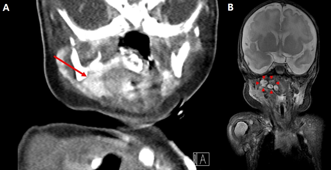
Figure 1. (A) A heterogeneous mass measuring 3.6×1.8×2 cm in size is located at the right oropharynx on computed tomography (arrow) and (B) composed of a tooth-like structure on magnetic resonance imaging (arrowheads).

Figure 2. (A, B) The right (Rt) condylar process was deviated, and the mandibular fossa demonstrated hypoplasia (arrows). (C) Facial computed tomography showed a subluxation of the temporomandibular joint (arrowheads). Abbreviation: Lt, left.

Figure 3. (A) A mass was seen in the right oropharyngeal region (arrow). (B) The oropharyngeal mass was then fully exposed, and part of the mass was removed (arrowheads). (C) Abnormalities of the tongue, such as a fissured tongue and accessory tongue, were observed, along with cleft palate (large arrow).

Figure 4. The oropharyngeal mass was whitish and round, measuring approximately 3.5×2.5×1.4 cm.
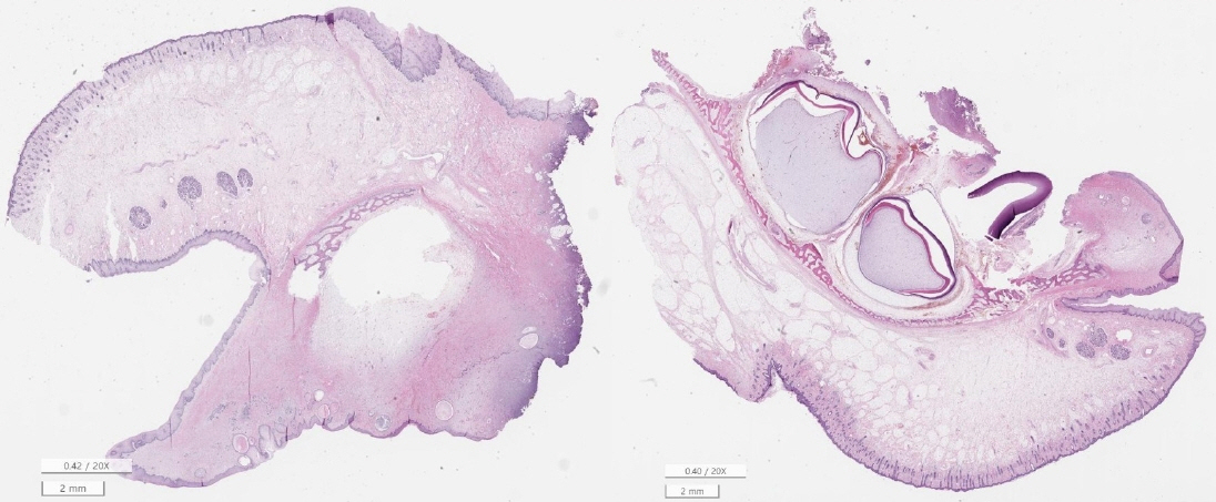
Figure 5. The biopsy results indicated the presence of a keratinizing squamous epithelial-lining lesion in the right oropharynx. It was eventually diagnosed as a mature teratoma (H&E stain, ×20).
Reference
-
1. Chiloiro S, Giampietro A, Bianchi A, De Marinis L. Clinical management of teratoma, a rare hypothalamic-pituitary neoplasia. Endocrine. 2016; 53:636–42.
Article2. Demajumdar R, Bhat N. Epignathus: a germ-cell tumour presenting as neonatal respiratory distress. Int J Pediatr Otorhinolaryngol. 1999; 47:87–90.
Article3. Acierno SP, Waldhausen JH. Congenital cervical cysts, sinuses and fistulae. Otolaryngol Clin North Am. 2007; 40:161–76.
Article4. Levine AB, Alvarez M, Wedgwood J, Berkowitz RL, Holzman I. Contemporary management of a potentially lethal fetal anomaly: a successful perinatal approach to epignathus. Obstet Gynecol. 1990; 76(5 Pt 2):962–6.5. Jayagobi PA, Chandran S, Sriram B, Chang KT. Ex-utero intrapartum treatment (EXIT) procedure for giant fetal epignathus. Indian Pediatr. 2015; 52:893–5.
Article6. Bale PM, Reye RD. Epignathus, double pituitary and agenesis of corpus callosum. J Pathol. 1976; 120:161–4.
Article7. April MM, Ward RF, Garelick JM. Diagnosis, management, and follow-up of congenital head and neck teratomas. Laryngoscope. 1998; 108:1398–401.
Article8. de Vries IA, Breugem CC, van der Heul AM, Eijkemans MJ, Kon M, Mink van der Molen AB. Prevalence of feeding disorders in children with cleft palate only: a retrospective study. Clin Oral Investig. 2014; 18:1507–15.
Article9. Enepekides DJ. Management of congenital anomalies of the neck. Facial Plast Surg Clin North Am. 2001; 9:131–45.10. Wiatrak BJ, Myer CM 3rd, Bratcher GO. Report of a nasopharyngeal teratoma evaluated with magnetic resonance imaging. Otolaryngol Head Neck Surg. 1990; 102:186–90.
Article11. Cil AS, Bozkurt M, Bozkurt DK. Intrauterine temporomandibular joint dislocation: prenatal sonographic evaluation. Clin Med Res. 2014; 12:58–60.
Article12. Okafor CI, Okafor CO, Odike MA. Congenital teratoma of the face. J Obstet Gynaecol. 2004; 24:828–9.
Article
- Full Text Links
-
- Actions
-
Cited
- CITED
-
- Close
- Share
-
- Similar articles
-
- A Case of Oropharyngeal Teratoma with Cleft Palate
- Chronic dislocation of temporomandibular joint persisting for 6 months: a case report
- Incidental finding of an extensive oropharyngeal mass in magnetic resonance imaging of a patient with temporomandibular disorder: A case report
- Re-restoration of temporomandibular joint disorder acquired after implant prosthetic restoration using T-Scan: A case report
- A study on simultation of the mandibular movement of the patients with temporomandibular joint disorder
