Restor Dent Endod.
2023 Aug;48(3):e27. 10.5395/rde.2023.48.e27.
Contemporary research trends on nanoparticles in endodontics: a bibliometric and scientometric analysis of the top 100 most-cited articles
- Affiliations
-
- 1Department of Endodontics, Gulhane Faculty of Dentistry, University of Health Sciences, Ankara, Turkey
- KMID: 2548230
- DOI: http://doi.org/10.5395/rde.2023.48.e27
Abstract
Objectives
Advancements in nanotechnology have led to the widespread usage of nanoparticles in the endodontic field. This bibliometric study aimed to determine and analyze the top 100 most-cited articles about nanoparticles in endodontics from 2000 to 2022.
Materials and Methods
A detailed electronic search was conducted on the “Clarivate Analytics Web of Science, All Databases” to receive the most-cited articles related to the topic. Articles were ranked in descending order based on their citation counts, and the first 100 were selected for bibliometric analysis. Parameters such as citation density, publication year, journal, country, institution, author, study design, study field, evidence level, and keywords were analyzed.
Results
The top 100 most-cited articles received 4,698 citations (16–271) with 970.21 (1.91– 181) citation density in total. Among decades, citations were significantly higher in 2011–2022 (p < 0.001). Journal of Endodontics had the largest number of publications. Canada and the University of Toronto made the highest contribution as country and institution, respectively. Anil Kishen was the 1 who participated in the largest number of articles. The majority of the articles were designed in vitro. The main study field was “antibacterial effect.” Among keywords, “nanoparticles” followed by “Enterococcus faecalis” were used more frequently.
Conclusions
Developments in nanotechnology had an impact on the increasing number of studies in recent years. This bibliometric study provides a comprehensive view of nanoparticle advances and trends using citation analysis.
Keyword
Figure
-
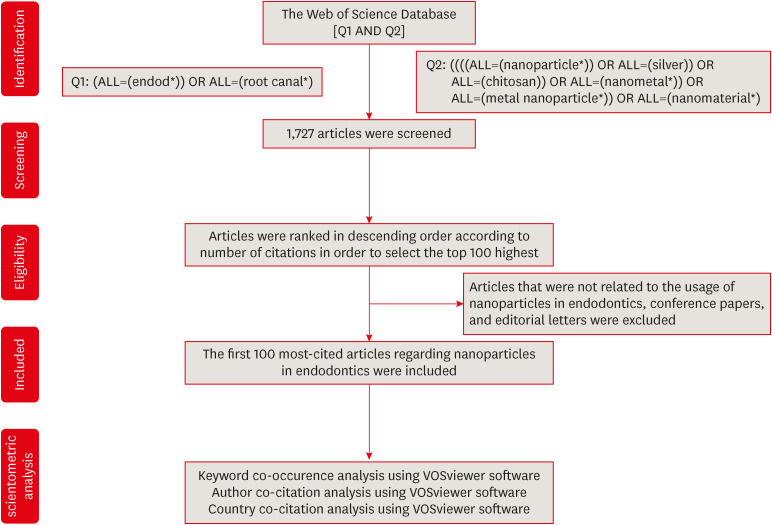
Figure 1 The flow diagram of the study.
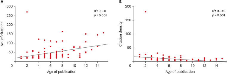
Figure 2 Association between the number of citations and age of publication (A), association between the citation density and age of publication (B).
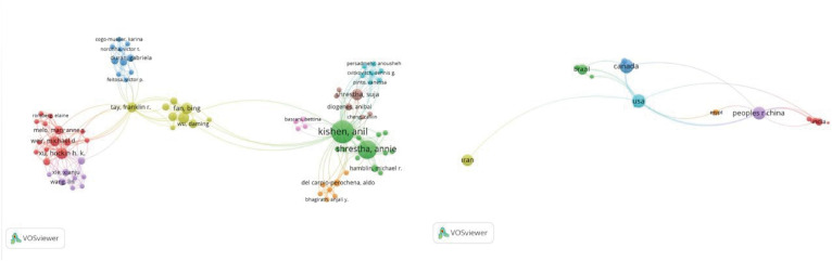
Figure 3 The visualization map of all contributed authors and countries to the articles about nanoparticles in endodontics, along with their collaboration.
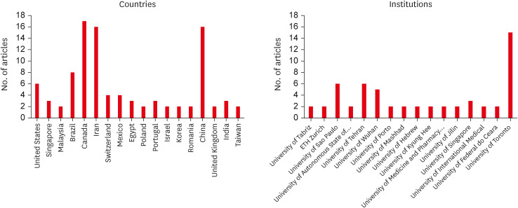
Figure 4 Participated countries and institutions with at least 2 publications.
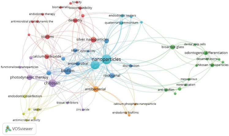
Figure 5 The visualization map of frequently used 2 or more co-occurring keywords of the top 100 most-cited articles about nanoparticles in endodontics.
Reference
-
1. European Commission. Commission recommendation of 18 October 2011 on the definition of nanomaterial (2011/696/EU). OJEU. 2011; 275:38–48.2. Schmalz G, Hickel R, van Landuyt KL, Reichl FX. Nanoparticles in dentistry. Dent Mater. 2017; 33:1298–1314. PMID: 28951037.
Article3. Bayda S, Adeel M, Tuccinardi T, Cordani M, Rizzolio F. The history of nanoscience and nanotechnology: from chemical-physical applications to nanomedicine. Molecules. 2019; 25:112. PMID: 31892180.
Article4. Shrestha A, Kishen A. Antibacterial nanoparticles in endodontics: a review. J Endod. 2016; 42:1417–1426. PMID: 27520408.5. Su M, Chen Y, Jia L, Zhang Z. Camptothecin-loaded and manganese dioxide-coated polydopamine nanomedicine used for magnetic resonance imaging diagnosis and chemo-photothermal therapy for lung cancer. Int J Nanomedicine. 2022; 17:6687–6705. PMID: 36597434.
Article6. Özdemir O, Kopac T. Recent progress on the applications of nanomaterials and nano-characterization techniques in endodontics: a review. Materials (Basel). 2022; 15:5109. PMID: 35897542.
Article7. Jandt KD, Watts DC. Nanotechnology in dentistry: present and future perspectives on dental nanomaterials. Dent Mater. 2020; 36:1365–1378. PMID: 32981749.
Article8. Wong J, Zou T, Lee AH, Zhang C. The potential translational applications of nanoparticles in endodontics. Int J Nanomedicine. 2021; 16:2087–2106. PMID: 33727815.
Article9. Fernandez CC, Sokolonski AR, Fonseca MS, Stanisic D, Araújo DB, Azevedo V, Portela RD, Tasic L. Applications of silver nanoparticles in dentistry: advances and technological innovation. Int J Mol Sci. 2021; 22:2485. PMID: 33801230.
Article10. Del Carpio-Perochena A, Kishen A, Felitti R, Bhagirath AY, Medapati MR, Lai C, Cunha RS. Antibacterial properties of chitosan nanoparticles and propolis associated with calcium hydroxide against single- and multispecies biofilms: an in vitro and in situ study. J Endod. 2017; 43:1332–1336. PMID: 28578886.
Article11. Raura N, Garg A, Arora A, Roma M. Nanoparticle technology and its implications in endodontics: a review. Biomater Res. 2020; 24:21. PMID: 33292702.
Article12. Afkhami F, Ahmadi P, Chiniforush N, Sooratgar A. Effect of different activations of silver nanoparticle irrigants on the elimination of Enterococcus faecalis . Clin Oral Investig. 2021; 25:6893–6899.
Article13. Zakrzewski W, Dobrzyński M, Zawadzka-Knefel A, Lubojański A, Dobrzyński W, Janecki M, Kurek K, Szymonowicz M, Wiglusz RJ, Rybak Z. Nanomaterials application in endodontics. Materials (Basel). 2021; 14:5296. PMID: 34576522.
Article14. Fan W, Wu D, Ma T, Fan B. Ag-loaded mesoporous bioactive glasses against Enterococcus faecalis biofilm in root canal of human teeth. Dent Mater J. 2015; 34:54–60. PMID: 25748459.
Article15. Upadya M, Shrestha A, Kishen A. Role of efflux pump inhibitors on the antibiofilm efficacy of calcium hydroxide, chitosan nanoparticles, and light-activated disinfection. J Endod. 2011; 37:1422–1426. PMID: 21924195.
Article16. Wu D, Fan W, Kishen A, Gutmann JL, Fan B. Evaluation of the antibacterial efficacy of silver nanoparticles against Enterococcus faecalis biofilm. J Endod. 2014; 40:285–290. PMID: 24461420.
Article17. Valderrama P, Baca P, Solana C, Ferrer-Luque CM. Root canal disinfection articles with the highest relative citation ratios. A bibliometric analysis from 1990 to 2019. Antibiotics (Basel). 2021; 10:1412. PMID: 34827350.
Article18. Aksoy S, Aksoy U, Orhan K. An overview of the 35 years of research in the oral radiology: a bibliometric analysis. Oral Radiol. 2022; 38:183–191. PMID: 34143357.
Article19. Adnan S, Ullah R. Top-cited articles in regenerative endodontics: a bibliometric analysis. J Endod. 2018; 44:1650–1664. PMID: 30243658.
Article20. Donthu N, Kumar S, Mukherjee D, Pandey N, Lim WM. How to conduct a bibliometric analysis: an overview and guidelines. J Bus Res. 2021; 133:285–296.
Article21. Shamszadeh S, Asgary S, Nosrat A. Regenerative endodontics: a scientometric and bibliometric analysis. J Endod. 2019; 45:272–280. PMID: 30803534.
Article22. de Araújo LP, da Rosa WL, Gobbo LB, da Silva TA, de Almeida JF, Ferraz CC. Global research trends on photodynamic therapy in endodontics: a bibliometric analysis. Photodiagnosis Photodyn Ther. 2022; 40:103039. PMID: 35907620.
Article23. Karobari MI, Maqbool M, Ahmad P, Abdul MS, Marya A, Venugopal A, Shaik GM, Scardina GA, Messina P, Noorani TY. Endodontic microbiology: a bibliometric analysis of the top 50 classics. BioMed Res Int. 2021; 2021:6657167. PMID: 34746305.
Article24. Silva EJ, Pinto KP, Ajuz NC, Sassone LM. Ten years of minimally invasive access cavities in Endodontics: a bibliometric analysis of the 25 most-cited studies. Restor Dent Endod. 2021; 46:e42. PMID: 34513648.
Article25. Kodonas K, Fardi A, Gogos C, Economides N. Top 50 cited articles on dental stem cell research. Restor Dent Endod. 2020; 45:e17. PMID: 32483534.
Article26. Murad MH, Asi N, Alsawas M, Alahdab F. New evidence pyramid. Evid Based Med. 2016; 21:125–127. PMID: 27339128.
Article27. Abou Neel EA, Bozec L, Perez RA, Kim HW, Knowles JC. Nanotechnology in dentistry: prevention, diagnosis, and therapy. Int J Nanomedicine. 2015; 10:6371–6394. PMID: 26504385.28. Waltman L, van Eck NJ, Wouters P. Counting publications and citations: is more always better? J Informetrics. 2013; 7:635–641.
Article29. Garfield E. Citation analysis as a tool in journal evaluation. Science. 1972; 178:471–479. PMID: 5079701.
Article30. Yin IX, Zhang J, Zhao IS, Mei ML, Li Q, Chu CH. The antibacterial mechanism of silver nanoparticles and its application in dentistry. Int J Nanomedicine. 2020; 15:2555–2562. PMID: 32368040.31. Waltimo T, Brunner TJ, Vollenweider M, Stark WJ, Zehnder M. Antimicrobial effect of nanometric bioactive glass 45S5. J Dent Res. 2007; 86:754–757. PMID: 17652205.
Article32. Yılmaz B, Dinçol ME, Yalçın TY. A bibliometric analysis of the 103 top-cited articles in endodontics. Acta Odontol Scand. 2019; 77:574–583. PMID: 31174442.
Article33. Kishen A, Shi Z, Shrestha A, Neoh KG. An investigation on the antibacterial and antibiofilm efficacy of cationic nanoparticulates for root canal disinfection. J Endod. 2008; 34:1515–1520. PMID: 19026885.
Article34. Shrestha A, Friedman S, Torneck CD, Kishen A. bioactivity of photoactivated functionalized nanoparticles assessed in lipopolysaccharide-contaminated root canals in vivo . J Endod. 2018; 44:104–110. PMID: 29153731.
Article35. Miranda R, Garcia-Carpintero E. Overcitation and overrepresentation of review papers in the most cited papers. J Informetrics. 2018; 12:1015–1030.
Article
- Full Text Links
-
- Actions
-
Cited
- CITED
-
- Close
- Share
-
- Similar articles
-
- The most influential articles on kidney transplantation: a bibliometric and visualized analysis
- The top 10 most-cited articles on the management of fractured instruments: a bibliometric analysis
- A bibliometric analysis on the most-cited publications on carotid endarterectomy throughout history
- Understanding anterior communicating artery aneurysms: A bibliometric analysis of top 100 most cited articles
- The Top-100 most cited articles on dural arteriovenous fistula: A bibliometric analysis
