Influence of access cavity design on calcium hydroxide removal using different cleaning protocols: a confocal laser scanning microscopy study
- Affiliations
-
- 1Department of Endodontics, School of Dentistry, İstanbul Health and Technology University, İstanbul, Turkey
- 2Department of Endodontics, School of Dentistry, Dicle University, Diyarbakır, Turkey
- 3Department of Endodontics, School of Dentistry, Eskişehir Osmangazi University, Eskişehir, Turkey
- 4Department of Endodontics, School of Dentistry, Universidade do Estado do Rio de Janeiro – UERJ, Rio de Janeiro, RJ, Brazil
- 5Department of Endodontics, School of Dentistry, İstanbul Medipol University, İstanbul, Turkey
- 6Department of Pedodontics, School of Dentistry, Afyonkarahisar Health Sciences University, Afyonkarahisar, Turkey
- KMID: 2548228
- DOI: http://doi.org/10.5395/rde.2023.48.e25
Abstract
Objectives
The purpose of this study was to evaluate the influence of endodontic access cavities design on the removal of calcium hydroxide medication of the apical third of mandibular incisor root canal walls and dentinal tubules with different cleaning protocols: EDDY sonic activation, Er,Cr:YSGG laser-activated irrigation, or conventional irrigation with IrriFlex.
Materials and Methods
Seventy-eight extracted human mandibular incisors were assigned to 6 experimental groups (n = 13) according to the endodontic access cavity and cleaning protocol for calcium hydroxide removal: traditional access cavity (TradAC)/EDDY; ultraconservative access cavity performed in the incisal edge (UltraAC.Inc)/EDDY; TradAC/Er,Cr:YSGG; UltraAC. Inc/Er,Cr:YSGG; TradAC/IrriFlex; or UltraAC.Inc/IrriFlex. Confocal laser scanning microscopy images were used to measure the non-penetration percentage, maximum residual calcium hydroxide penetration depth, and penetration area at 2 and 4 mm from the apex. Data were statistically analyzed using Shapiro-Wilk and WRS2 package for 2-way comparison of nonnormally distributed parameters (depth of penetration, area of penetration, and percentage of non-penetration) according to cavity and cleaning protocol with the significance level set at 5%.
Results
The effect of cavity and cleaning protocol interactions on penetration depth, penetration area and non-penetration percentage was not found statistically significant at 2 and 4 mm levels (p > 0.05).
Conclusions
The present study demonstrated that TradAC or UltraAC.Inc preparations with different cleaning protocols in extracted mandibular incisors did not influence the remaining calcium hydroxide at 2 and 4 mm from the apex.
Figure
-
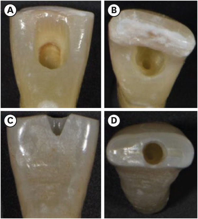
Figure 1 Photographs demonstrating the cavity designs. (A, B) Traditional access cavity and (C, D) ultraconservative access cavity performed in the incisal edge.
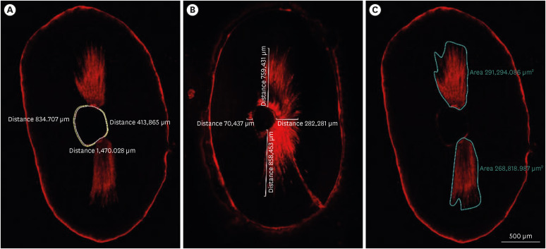
Figure 2 Image analyses procedures. (A) The non-penetration percentage, (B) maximum residual calcium hydroxide penetration depth, and (C) penetration area of the calcium hydroxide inside the dentinal tubules.
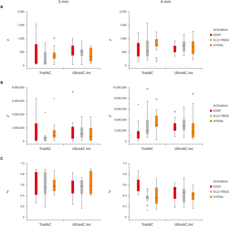
Figure 3 Box plot presentation of (A) penetration depth, (B) penetration area, and (C) non-penetration percentages measured at 2 and 4 mm levels from the apex for comparing TradAC and UltraAC.Inc with different cleaning protocols.TradAC, traditional access cavity; UltraAC.Inc, ultraconservative access cavity performed in the incisal edge.
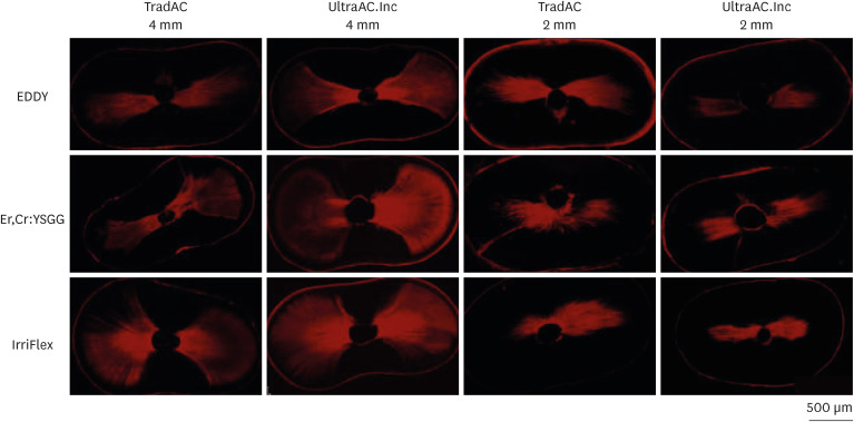
Figure 4 Confocal laser scanning microscopy representative images of the calcium hydroxide penetration of TradAC and UltraAC.Inc groups in the apical root third (4 and 2 mm) of specimens. The effect of access cavity preparation and cleaning protocols was not found statistically significant for both 2 and 4 mm level.TradAC, traditional access cavity; UltraAC.Inc, ultraconservative access cavity performed in the incisal edge.
Reference
-
1. Ingle JI, Walton RE, Lambert GL, Lambert C, Taintor JF, Zidell JD, Beveridge EE. Preparation for endodontic therapy. Ingle JI, Taintor JF, editors. Endodontics. 3rd ed. Philadelphia, PA: Lea & Febiger;1985. p. 54–101.2. Korzen BH, Pulver WH. Endodontic access cavities--the first step to success. Ont Dent. 1978; 55:19–22.3. LaTurno SA, Zillich RM. Straight-line endodontic access to anterior teeth. Oral Surg Oral Med Oral Pathol. 1985; 59:418–419. PMID: 3858780.
Article4. Levin HJ. Access cavities. Dent Clin North Am. 1967; 11:701–710.
Article5. Silva AA, Belladonna FG, Rover G, Lopes RT, Moreira EJ, De-Deus G, Silva EJ. Does ultraconservative access affect the efficacy of root canal treatment and the fracture resistance of two-rooted maxillary premolars? Int Endod J. 2020; 53:265–275. PMID: 31519039.
Article6. Silva EJ, De-Deus G, Souza EM, Belladonna FG, Cavalcante DM, Simões-Carvalho M, Versiani MA. Present status and future directions - Minimal endodontic access cavities. Int Endod J. 2022; 55(Suppl 3):531–587.
Article7. Vieira GC, Pérez AR, Alves FR, Provenzano JC, Mdala I, Siqueira JF Jr, Rôças IN. Impact of contracted endodontic cavities on root canal disinfection and shaping. J Endod. 2020; 46:655–661. PMID: 32201072.
Article8. Silva EJ, Pinto KP, Ferreira CM, Belladonna FG, De-Deus G, Dummer PM, Versiani MA. Current status on minimal access cavity preparations: a critical analysis and a proposal for a universal nomenclature. Int Endod J. 2020; 53:1618–1635. PMID: 32854167.
Article9. Abou-Elnaga MY, Alkhawas MA, Kim HC, Refai AS. Effect of truss access and artificial truss restoration on the fracture resistance of endodontically treated mandibular first molars. J Endod. 2019; 45:813–817. PMID: 30905571.
Article10. Santosh SS, Ballal S, Natanasabapathy V. Influence of minimally invasive access cavity designs on the fracture resistance of endodontically treated mandibular molars subjected to thermocycling and dynamic loading. J Endod. 2021; 47:1496–1500. PMID: 34237385.
Article11. Augusto CM, Barbosa AF, Guimarães CC, Lima CO, Ferreira CM, Sassone LM, Silva EJ. A laboratory study of the impact of ultraconservative access cavities and minimal root canal tapers on the ability to shape canals in extracted mandibular molars and their fracture resistance. Int Endod J. 2020; 53:1516–1529. PMID: 32683704.
Article12. Barbosa AF, Silva EJ, Coelho BP, Ferreira CM, Lima CO, Sassone LM. The influence of endodontic access cavity design on the efficacy of canal instrumentation, microbial reduction, root canal filling and fracture resistance in mandibular molars. Int Endod J. 2020; 53:1666–1679. PMID: 32762041.
Article13. Maske A, Weschenfelder VM, Soares Grecca Vilella F, Burnett Junior LH, de Melo TA. Influence of access cavity design on fracture strength of endodontically treated lower molars. Aust Endod J. 2021; 47:5–10. PMID: 32981120.
Article14. Kim D, Kim E. Antimicrobial effect of calcium hydroxide as an intracanal medicament in root canal treatment: a literature review - Part II. in vivo studies. Restor Dent Endod. 2015; 40:97–103. PMID: 25984470.
Article15. Taşdemir T, Çelik D, Er K, Yildirim T, Ceyhanli KT, Yeşilyurt C. Efficacy of several techniques for the removal of calcium hydroxide medicament from root canals. Int Endod J. 2011; 44:505–509. PMID: 21276018.
Article16. Capar ID, Ozcan E, Arslan H, Ertas H, Aydinbelge HA. Effect of different final irrigation methods on the removal of calcium hydroxide from an artificial standardized groove in the apical third of root canals. J Endod. 2014; 40:451–454. PMID: 24565670.
Article17. Kim SK, Kim YO. Influence of calcium hydroxide intracanal medication on apical seal. Int Endod J. 2002; 35:623–628. PMID: 12190902.
Article18. Lambrianidis T, Kosti E, Boutsioukis C, Mazinis M. Removal efficacy of various calcium hydroxide/chlorhexidine medicaments from the root canal. Int Endod J. 2006; 39:55–61. PMID: 16409329.
Article19. Innovative Sonic Powered Irrigation. cited March 1, 2023. Available from: https://www.vdw-dental.com/.20. Donnermeyer D, Wyrsch H, Bürklein S, Schäfer E. Removal of calcium hydroxide from artificial grooves in straight root canals: sonic activation using eddy versus passive ultrasonic irrigation and XPendo finisher. J Endod. 2019; 45:322–326. PMID: 30803540.
Article21. Marques-da-Silva B, Alberton CS, Tomazinho FS, Gabardo MC, Duarte MA, Vivan RR, Baratto-Filho F. Effectiveness of five instruments when removing calcium hydroxide paste from simulated internal root resorption cavities in extracted maxillary central incisors. Int Endod J. 2020; 53:366–375. PMID: 31566756.
Article22. Güven Y, Ali A, Arslan H. Efficiency of Endosonic Blue, EDDY, Ultra X and Endoactivator in the removal of calcium hydroxide paste from root canals. Aust Endod J. 2022; 48:32–36. PMID: 34939722.
Article23. Blanken J, Verdaasdonk R. Cavitation as a working mechanism of the Er,Cr:YSGG laser in endodontics: a visualization study. J Oral Laser Appl. 2007; 7:97–106.24. de Groot SD, Verhaagen B, Versluis M, Wu MK, Wesselink PR, van der Sluis LW. Laser-activated irrigation within root canals: cleaning efficacy and flow visualization. Int Endod J. 2009; 42:1077–1083. PMID: 19912378.
Article25. DiVito E, Peters OA, Olivi G. Effectiveness of the erbium:YAG laser and new design radial and stripped tips in removing the smear layer after root canal instrumentation. Lasers Med Sci. 2012; 27:273–280. PMID: 21120568.
Article26. Eymirli A, Nagas E, Uyanik MO, Cehreli ZC. Effect of laser-activated ırrigation with ethylene diaminetetraacetic acid and phytic acid on the removal of calcium hydroxide and triple antibiotic paste from root dentin. Photomed Laser Surg. 2017; 35:43–48. PMID: 27623238.
Article27. IrriFlex. IrriFlex brochure. cited July 6, 2022. Available from: https://pd-irriflex.com/.28. Krishan R, Paqué F, Ossareh A, Kishen A, Dao T, Friedman S. Impacts of conservative endodontic cavity on root canal instrumentation efficacy and resistance to fracture assessed in incisors, premolars, and molars. J Endod. 2014; 40:1160–1166. PMID: 25069925.
Article29. Dias-Junior LCL, Castro RF, Fernandes AD, Guerreiro MYR, Silva EJNL, Brandão JMDS. Final endodontic irrigation with 70% ethanol enhanced calcium hydroxide removal from the apical third. J Endod. 2021; 47:105–111. PMID: 33045271.
Article30. Abduljalil M, Kalender A. Efficacy of Er,Cr:YSGG laser with different output powers on removing smear layer after retreatment of two different obturation techniques. Photobiomodul Photomed Laser Surg. 2020; 38:84–90. PMID: 31339812.
Article31. Moon YM, Shon WJ, Baek SH, Bae KS, Kum KY, Lee W. Effect of final irrigation regimen on sealer penetration in curved root canals. J Endod. 2010; 36:732–736. PMID: 20307754.
Article32. Mair P, Wilcox R. Robust statistical methods in R using the WRS2 package. Behav Res Methods. 2020; 52:464–488. PMID: 31152384.
Article33. Yaylali IE, Kececi AD, Ureyen Kaya B. Ultrasonically activated irrigation to remove calcium hydroxide from apical third of human root canal system: a systematic review of in vitro studies. J Endod. 2015; 41:1589–1599. PMID: 26238527.34. Gokturk H, Ozkocak I, Buyukgebiz F, Demir O. Effectiveness of various irrigation protocols for the removal of calcium hydroxide from artificial standardized grooves. J Appl Oral Sci. 2017; 25:290–298. PMID: 28678948.
Article35. Suresh N, Varghese A, Sundar S, Nagendrababu V, Velmurugan N. Do calcium chelators play a role in the removal of calcium hydroxide from root canals? A systematic review of laboratory studies. Eur Endod J. 2022; 7:11–19. PMID: 35353065.
Article36. Küçükkaya Eren S, Uzunoĝlu Özyürek E. Influence of cavity design on calcium hydroxide removal from root canal irregularities. Cumhur Dent J. 2019; 22:2146–2852.37. Silva LJ, Pessoa OF, Teixeira MB, Gouveia CH, Braga RR. Micro-CT evaluation of calcium hydroxide removal through passive ultrasonic irrigation associated with or without an additional instrument. Int Endod J. 2015; 48:768–773. PMID: 25156123.
Article38. Komabayashi T, D’souza RN, Dechow PC, Safavi KE, Spångberg LS. Particle size and shape of calcium hydroxide. J Endod. 2009; 35:284–287. PMID: 19166791.
Article39. Deniz Sungur D, Purali N, Coşgun E, Calt S. Push-out bond strength and dentinal tubule penetration of different root canal sealers used with coated core materials. Restor Dent Endod. 2016; 41:114–120. PMID: 27200279.
Article40. Carrigan PJ, Morse DR, Furst ML, Sinai IH. A scanning electron microscopic evaluation of human dentinal tubules according to age and location. J Endod. 1984; 10:359–363. PMID: 6590745.
Article41. Rover G, de Lima CO, Belladonna FG, Garcia LF, Bortoluzzi EA, Silva EJ, Teixeira CS. Influence of minimally invasive endodontic access cavities on root canal shaping and filling ability, pulp chamber cleaning and fracture resistance of extracted human mandibular incisors. Int Endod J. 2020; 53:1530–1539. PMID: 32754937.
Article42. Rödig T, Koberg C, Baxter S, Konietschke F, Wiegand A, Rizk M. Micro-CT evaluation of sonically and ultrasonically activated irrigation on the removal of hard-tissue debris from isthmus-containing mesial root canal systems of mandibular molars. Int Endod J. 2019; 52:1173–1181. PMID: 30773661.
Article43. Blanken J, De Moor RJ, Meire M, Verdaasdonk R. Laser induced explosive vapor and cavitation resulting in effective irrigation of the root canal. Part 1: a visualization study. Lasers Surg Med. 2009; 41:514–519. PMID: 19639622.
Article44. Yavari HR, Rahimi S, Shahi S, Lotfi M, Barhaghi MH, Fatemi A, Abdolrahimi M. Effect of Er,Cr:YSGG laser irradiation on Enterococcus faecalis in infected root canals. Photomed Laser Surg. 2010; 28(Suppl 1):S91–S96. PMID: 20666572.45. Kuştarcı A, Er K, Siso SH, Aydın H, Harorlı H, Arslan D, Kirmali O. Efficacy of laser-activated irrigants in calcium hydroxide removal from the artificial grooves in root canals: an ex vivo study. Photomed Laser Surg. 2016; 34:205–210. PMID: 27058651.
Article46. Silva EJ, Versiani MA, Souza EM, De-Deus G. Minimally invasive access cavities: does size really matter? Int Endod J. 2021; 54:153–155. PMID: 33452846.
Article47. Gulabivala K, Ng YL. Non-surgical root-canal treatment. Endodontics. 4th ed. Edinburgh: Mosby/Elsevier Ltd.;2014. p. 174–236.
- Full Text Links
-
- Actions
-
Cited
- CITED
-
- Close
- Share
-
- Similar articles
-
- The Change of Epidermal Calcium Gradient: Confocal Laser Scanning Microscopic Approach
- Bonding effects of cleaning protocols and time-point of acid etching on dentin impregnated with endodontic sealer
- Application of Autofluorescence for Confocal Microscopy to Aid in Archaeoparasitological Analyses
- Intravital Laser-scanning Two-photon and Confocal Microscopy for Biomedical Research
- Effect of calcium hydroxide application time on dentin
