Restor Dent Endod.
2023 Aug;48(3):e23. 10.5395/rde.2023.48.e23.
Radiographic patterns of periosteal bone reactions associated with endodontic lesions
- Affiliations
-
- 1Department of Endodontics, Texas A&M University (TAMU) School of Dentistry, Dallas, TX, USA
- 2Department of Diagnostic Sciences, Texas A&M University (TAMU) School of Dentistry, Dallas, TX, USA
- KMID: 2548226
- DOI: http://doi.org/10.5395/rde.2023.48.e23
Abstract
Objectives
The formation of new bone by periosteum due to an insult is called periosteal bone reaction (PBR). This study assessed the cone beam computed tomography (CBCT) patterns of periosteal bone reactions associated with periapical inflammatory lesion (apical periodontitis/periapical rarefying osteitis).
Materials and Methods
Twenty-two small field of view CBCT images of patients with PBR were selected from a database of a private practice limited to endodontics. The volume of the periapical inflammatory lesion, the presence of cortical fenestration, the distance of the root apices to the affected cortex, and the location, pattern, and longest diameter of the periosteal reaction were recorded. Statistical analysis was performed using Wilcoxon Ranksum, Fischer’s exact, Spearman Correlation Coefficient, and paired t-test.
Results
In all cases, periosteal bone reaction manifested as either parallel (90.9%) or irregular (9.1%). No correlation was found between periapical inflammatory lesion volume and the periosteal reaction's longest diameter (p > 0.05). Cortical fenestration was noted in 72.7% of the cases. In addition, the findings showed that periosteal reactions were located mostly on the buccal and were present 53.8% and 100% of the time in the mandible and maxilla, respectively.
Conclusions
The periosteal reactions of endodontic origin had a nonaggressive form (i.e., parallel or irregular), and none of the lesions resulted in a periosteal reaction with an ominous Codman’s triangle or spicule pattern.
Keyword
Figure
-

Figure 1 The volume of the apical periodontitis/periapical rarefying osteitis was calculated using Mimics Innovation Suite.
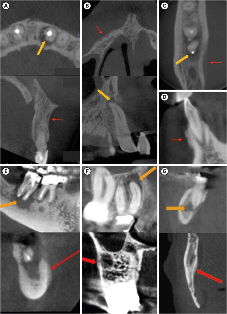
Figure 2 Cone beam computed tomography images of 7 cases with periosteal reactions (red arrow) and periapical inflammatory lesion (yellow arrow). (A-C), (E-G) manifesting parallel periosteal reactions, and (D) showing irregular periosteal reaction.
Reference
-
1. Chen EM, Masih S, Chow K, Matcuk G, Patel D. Periosteal reaction: review of various patterns associated with specific pathology. Contemp Diagn Radiol. 2012; 35:1–5.2. Rana RS, Wu JS, Eisenberg RL. Periosteal reaction. AJR Am J Roentgenol. 2009; 193:W259-72. PMID: 19770293.
Article3. Seok H, Kim SG, Song JY. Proliferative periostitis of the mandibular ramus and condyle: a case report. J Korean Assoc Oral Maxillofac Surg. 2015; 41:198–202. PMID: 26339579.
Article4. Koorbusch GF, Deatherage JR, Curé JK. How can we diagnose and treat osteomyelitis of the jaws as early as possible? Oral Maxillofac Surg Clin North Am. 2011; 23:557–567. PMID: 21982609.
Article5. Ida M, Tetsumura A, Kurabayashi T, Sasaki T. Periosteal new bone formation in the jaws. A computed tomographic study. Dentomaxillofac Radiol. 1997; 26:169–176. PMID: 9442603.
Article6. Jalali P, Tahmasbi M, Augsburger RA, Khalilkhani NK, Daghighi K. Dynamics of bone loss in cases with acute or chronic apical abscess. J Endod. 2019; 45:1114–1118. PMID: 31280911.
Article7. Ida M, Watanabe H, Tetsumura A, Kurabayashi T. CT findings as a significant predictive factor for the curability of mandibular osteomyelitis: multivariate analysis. Dentomaxillofac Radiol. 2005; 34:86–90. PMID: 15829690.
Article8. Hayashi Y, Shimizutani K, Koseki Y. Radiographic study of periosteal new bone formation in osteomyelitis of the mandible. Oral Radiol. 1992; 8:19–26.
Article9. Bougatef H, Moussaoui E, Kallel I, Smaoui M, Oualha L, Douki N. Periostitis ossificans: report of two cases resolved with endodontic treatment. Case Rep Dent. 2020; 2020:8876268. PMID: 33299618.
Article10. Zand V, Lotfi M, Vosoughhosseini S. Proliferative periostitis: a case report. J Endod. 2008; 34:481–483. PMID: 18358903.
Article11. de Andrade Freitas Oliveira LS, de Oliveira TFL, de Melo DP, de Menezes AV, Crusoé-Rebello I, Campos PSF. Computed Tomography findings of periostitis ossificans. Braz J Oral Sci. 2010; 9:59–62.
- Full Text Links
-
- Actions
-
Cited
- CITED
-
- Close
- Share
-
- Similar articles
-
- Computed tomography analysis of primary bone tumors : the significance in the evaluation of destructive lesionsof pelvic bone
- Periosteal Reaction of Osteomyelitis: MRI Findings Compared with Plain Radiographs
- A lamellar nodular periosteal reaction occuring in osteomyelitis: useful finding for differentiation from malignant bone tumor
- Osteosarcoma arising on the surface of long bones: a review of radiologic and histologic spectrum in 9 cases
- Study of Periosteal Reaction in Normal Infants
