Restor Dent Endod.
2023 May;48(2):e17. 10.5395/rde.2023.48.e17.
Successful nonsurgical treatment of type II dens invaginatus with 5 root canals using a self-adjusting file: a case report
- Affiliations
-
- 1Postgraduate Program in Dental Sciences, University Center Christus, Fortaleza, CE, Brazil
- 2Postgraduate Program in Dentistry, Estácio de Sá University, Rio de Janeiro, RJ, Brazil
- 3Postgraduate Program in Dentistry, University of Grande Rio (UNIGRANRIO), Duque de Caxias, RJ, Brazil
- 4Department of Endodontics and Dental Research Group, Iguaçu University (UNIG), Nova Iguaçu, RJ, Brazil
- KMID: 2548220
- DOI: http://doi.org/10.5395/rde.2023.48.e17
Abstract
- The present report describes the endodontic treatment of an Oehlers type II dens invaginatus in a maxillary lateral incisor with 5 root canals, an extremely rare condition. Apical periodontitis and related symptoms were noted. Cone-beam computed tomography was used to aid the diagnosis, reveal tooth morphology, and assist in canal location. The pulp chamber was carefully accessed, and the root canals were explored under magnification. All root canals were prepared with an R25 Reciproc Blue system and sodium hypochlorite (NaOCl) irrigation. After initial preparation, a self-adjusting file (SAF) with NaOCl and ethylenediaminetetraacetic acid was used to complement the disinfection. Additionally, calcium hydroxide medication was applied. Vertical compaction was used to fill the canals with a calcium silicate-based endodontic sealer and gutta-percha. After 12 months, the patient exhibited healing of the periapical region, absence of symptoms, and normal dental function. In conclusion, this nonsurgical treatment protocol was successful in promoting the cure of apical periodontitis. Both complementary disinfection with an SAF and use of calcium hydroxide medication should be considered when choosing the best treatment approach for dens invaginatus with very complex anatomy.
Figure
-

Figure 1 Clinical aspect of the tooth crown and initial radiographic image.
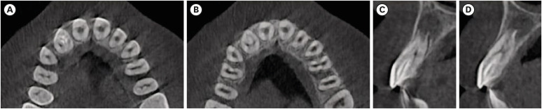
Figure 2 Cone-beam computed tomographic images.

Figure 3 Access cavity including both the true root canal and the invagination and root canal exploration (clinical and radiographic views).
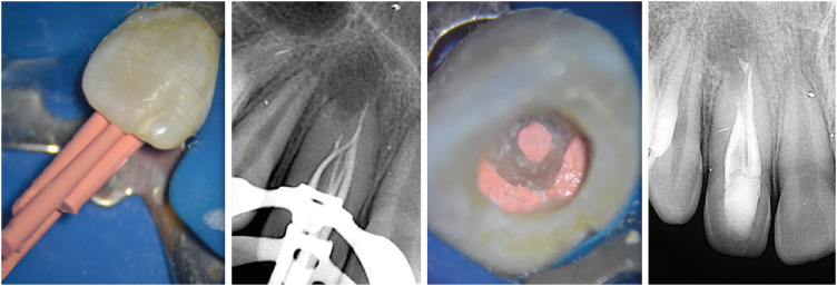
Figure 4 Details of the filling phase.
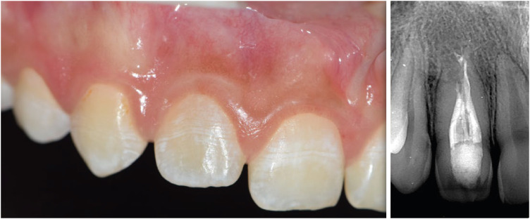
Figure 5 Clinical and radiographic aspects 2 years after treatment.
Reference
-
1. Wolcott J, Ishley D, Kennedy W, Johnson S, Minnich S, Meyers J. A 5 yr clinical investigation of second mesiobuccal canals in endodontically treated and retreated maxillary molars. J Endod. 2005; 31:262–264. PMID: 15793380.
Article2. Song M, Kim HC, Lee W, Kim E. Analysis of the cause of failure in nonsurgical endodontic treatment by microscopic inspection during endodontic microsurgery. J Endod. 2011; 37:1516–1519. PMID: 22000454.
Article3. Nosrat A, Schneider SC. Endodontic management of a maxillary lateral incisor with 4 root canals and a dens invaginatus tract. J Endod. 2015; 41:1167–1171. PMID: 25799535.
Article4. Oehlers FA. Dens invaginatus (dilated composite odontome). I. Variations of the invagination process and associated anterior crown forms. Oral Surg Oral Med Oral Pathol. 1957; 10:1204–1218 contd. PMID: 13477660.5. Rotstein I, Stabholz A, Heling I, Friedman S. Clinical considerations in the treatment of dens invaginatus. Endod Dent Traumatol. 1987; 3:249–254. PMID: 3479324.
Article6. Gonçalves A, Gonçalves M, Oliveira DP, Gonçalves N. Dens invaginatus type III: report of a case and 10-year radiographic follow-up. Int Endod J. 2002; 35:873–879. PMID: 12406383.
Article7. Alani A, Bishop K. Dens invaginatus. Part 1: classification, prevalence and aetiology. Int Endod J. 2008; 41:1123–1136. PMID: 19133103.
Article8. Metzger Z, Teperovich E, Zary R, Cohen R, Hof R. The self-adjusting file (SAF). Part 1: respecting the root canal anatomy--a new concept of endodontic files and its implementation. J Endod. 2010; 36:679–690. PMID: 20307744.
Article9. Matherne RP, Angelopoulos C, Kulild JC, Tira D. Use of cone-beam computed tomography to identify root canal systems in vitro . J Endod. 2008; 34:87–89. PMID: 18155501.
Article10. Nagendrababu V, Chong BS, McCabe P, Shah PK, Priya E, Jayaraman J, Pulikkotil SJ, Setzer FC, Sunde PT, Dummer PM. PRICE 2020 guidelines for reporting case reports in endodontics: a consensus-based development. Int Endod J. 2020; 53:619–626. PMID: 32090342.
Article11. Capar ID, Ertas H, Arslan H, Tarim Ertas E. A retrospective comparative study of cone-beam computed tomography versus rendered panoramic images in identifying the presence, types, and characteristics of dens invaginatus in a Turkish population. J Endod. 2015; 41:473–478. PMID: 25595467.
Article12. Koteeswaran V, Chandrasekaran S, Natanasabapathy V. Endodontic management of double dens invaginatus in maxillary central incisor. J Conserv Dent. 2018; 21:574–577. PMID: 30294124.
Article13. Bishop K, Alani A. Dens invaginatus. Part 2: clinical, radiographic features and management options. Int Endod J. 2008; 41:1137–1154. PMID: 19133104.
Article14. Metzger Z. The self-adjusting file (SAF) system: an evidence-based update. J Conserv Dent. 2014; 17:401–419. PMID: 25298639.
Article15. Siqueira JF Jr, Alves FR, Almeida BM, de Oliveira JC, Rôças IN. Ability of chemomechanical preparation with either rotary instruments or self-adjusting file to disinfect oval-shaped root canals. J Endod. 2010; 36:1860–1865. PMID: 20951301.
Article16. Paqué F, Peters OA. Micro-computed tomography evaluation of the preparation of long oval root canals in mandibular molars with the self-adjusting file. J Endod. 2011; 37:517–521. PMID: 21419301.
Article17. Metzger Z, Teperovich E, Cohen R, Zary R, Paqué F, Hülsmann M. The self-adjusting file (SAF). Part 3: removal of debris and smear layer-a scanning electron microscope study. J Endod. 2010; 36:697–702. PMID: 20307746.
Article18. Rôças IN, Provenzano JC, Neves MS, Alves FR, Gonçalves LS, Siqueira JF Jr. Effects of calcium hydroxide paste in different vehicles on bacterial reduction during treatment of teeth with apical periodontitis. J Endod. 2023; 49:55–61. PMID: 36309246.
Article19. Vera J, Siqueira JF Jr, Ricucci D, Loghin S, Fernández N, Flores B, Cruz AG. One- versus two-visit endodontic treatment of teeth with apical periodontitis: a histobacteriologic study. J Endod. 2012; 38:1040–1052. PMID: 22794203.
Article20. Leonardo MR, Almeida WA, Ito IY, da Silva LA. Radiographic and microbiologic evaluation of posttreatment apical and periapical repair of root canals of dogs’ teeth with experimentally induced chronic lesion. Oral Surg Oral Med Oral Pathol. 1994; 78:232–238. PMID: 7936595.
Article21. Hernández SR, Pérez AR, Baasch AC, Brisson-Suárez K, Sellera DP, Ferrari PH, Alberdi JC, Carreira CM, Gomes-Azevedo S, Alves FR, Rôças IN, Siqueira JF Jr. Management of teeth with dens invaginatus and apical periodontitis: a case series. J Am Dent Assoc. 2022; 153:470–478. PMID: 35184866.22. De Rossi A, Carvalho FK, Queiróz AM, Silva RA, Nelson-Filho P, Silva LA. The treatment of a maxillary lateral incisor with unusual morphology with long-term follow-up. Int Endod J. 2013; 46:1183–1190. PMID: 23594115.
Article23. Jaramillo A, Fernández R, Villa P. Endodontic treatment of dens invaginatus: a 5-year follow-up. Oral Surg Oral Med Oral Pathol Oral Radiol Endod. 2006; 101:e15–e21. PMID: 16360600.
Article24. Sundqvist G, Figdor D, Persson S, Sjögren U. Microbiologic analysis of teeth with failed endodontic treatment and the outcome of conservative re-treatment. Oral Surg Oral Med Oral Pathol Oral Radiol Endod. 1998; 85:86–93. PMID: 9474621.
Article
- Full Text Links
-
- Actions
-
Cited
- CITED
-
- Close
- Share
-
- Similar articles
-
- Root canal therapy of anterior teeth with dens invaginatus
- Endodontic management of a maxillary lateral incisor with dens invaginatus and external root irregularity using cone-beam computed tomography
- Guided endodontics: a case report of maxillary lateral incisors with multiple dens invaginatus
- A comparison of master apical file size according to instrumentation in type II root canal
- A case report of multiple bilateral dens invaginatus in maxillary anteriors
