Influence of CBCT parameters on image quality and the diagnosis of vertical root fractures in teeth with metallic posts: an ex vivo study
- Affiliations
-
- 1Department of Oral Diagnosis, Division of Oral Radiology, Piracicaba Dental School, University of Campinas, Piracicaba, São Paulo, Brazil
- 2Dental Clinic Department, Division of Oral Radiology, Dental School, Federal Fluminense University, Niterói, Rio de Janeiro, Brazil
- 3Department of Diagnosis and Oral Health, University of Louisville, School of Dentistry, Louisville, KY, USA
- KMID: 2548219
- DOI: http://doi.org/10.5395/rde.2023.48.e16
Abstract
Objectives
The aim of this study was to evaluate the influence of peak kilovoltage (kVp) and a metal artifact reduction (MAR) tool on image quality and the diagnosis of vertical root fracture (VRF) in cone-beam computed tomography (CBCT).
Materials and Methods
Twenty single-rooted human teeth filled with an intracanal metal post were divided into 2 groups: control (n = 10) and VRF (n = 10). Each tooth was placed into the socket of a dry mandible, and CBCT scans were acquired using a Picasso Trio varying the kVp (70, 80, 90, or 99), and the use of MAR (with or without). The examinations were assessed by 5 examiners for the diagnosis of VRF using a 5-point scale. A subjective evaluation of the expression of artifacts was done by comparing random axial images of the studied protocols. The results of the diagnoses were analyzed using 2-way analysis of variance and the Tukey post hoc test, the subjective evaluations were compared using the Friedman test, and intra-examiner reproducibility was evaluated using the weighted kappa test (α = 5%).
Results
The kVp and MAR did not influence the diagnosis of VRF (p > 0.05). According to the subjective classification, the 99 kVp protocol with MAR demonstrated the least expression of artifacts, while the 70 kVp protocol without MAR led to the most artifacts.
Conclusions
Protocols with higher kVp combined with MAR improved the image quality of CBCT examinations. However, those factors did not lead to an improvement in the diagnosis of VRF.
Figure
-
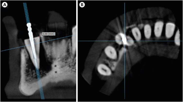
Figure 1 Obtaining the axial images used in the qualitative assessment of artifacts in all protocols. (A) Axial orientation line positioned 3 mm from the superior limit of the root and perpendicular to the root in an adjusted coronal reconstruction. (B) Axial reconstruction used in the template.
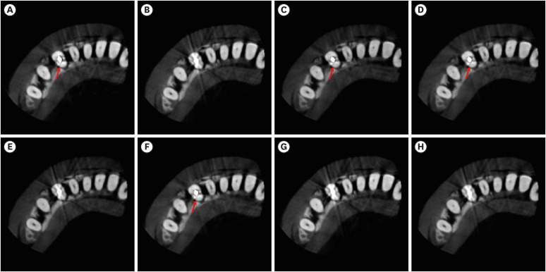
Figure 2 Example of the template used in the qualitative evaluation of artifacts. The images correspond to the axial reconstructions of the same tooth (of the group with vertical root fracture) obtained with all the studied protocols. (A) 70 kVp with MAR. (B) 80 kVp without MAR. (C) 99 kVp with MAR. (D) 90 kVp with MAR. (E) 99 kVp without MAR. (F) 80 kVp with MAR. (G) 70 kVp without MAR. (H) 90 kVp without MAR. The arrows indicate the fracture.kVp, peak kilovoltage; MAR, metal artifact reduction.
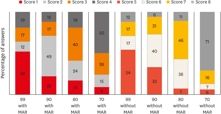
Figure 3 Distribution of the responses of the examiners for artifact expression in all protocols.kVp, peak kilovoltage; MAR, metal artifact reduction.
Reference
-
1. Ferreira LM, Visconti MA, Nascimento HA, Dallemolle RR, Ambrosano GM, Freitas DQ. Influence of CBCT enhancement filters on diagnosis of vertical root fractures: a simulation study in endodontically treated teeth with and without intracanal posts. Dentomaxillofac Radiol. 2015; 44:20140352. PMID: 25666446.
Article2. Yamamoto-Silva FP, de Oliveira Siqueira CF, Silva MA, Fonseca RB, Santos AA, Estrela C, de Freitas Silva BS. Influence of voxel size on cone-beam computed tomography-based detection of vertical root fractures in the presence of intracanal metallic posts. Imaging Sci Dent. 2018; 48:177–184. PMID: 30276154.
Article3. Chang E, Lam E, Shah P, Azarpazhooh A. Cone-beam computed tomography for detecting vertical root fractures in endodontically treated teeth: a systematic review. J Endod. 2016; 42:177–185. PMID: 26631300.
Article4. Freitas DQ, Vasconcelos TV, Noujeim M. Diagnosis of vertical root fracture in teeth close and distant to implant: an in vitro study to assess the influence of artifacts produced in cone beam computed tomography. Clin Oral Investig. 2019; 23:1263–1270.
Article5. Schulze R, Heil U, Gross D, Bruellmann DD, Dranischnikow E, Schwanecke U, Schoemer E. Artefacts in CBCT: a review. Dentomaxillofac Radiol. 2011; 40:265–273. PMID: 21697151.
Article6. Queiroz PM, Oliveira ML, Groppo FC, Haiter-Neto F, Freitas DQ. Evaluation of metal artefact reduction in cone-beam computed tomography images of different dental materials. Clin Oral Investig. 2018; 22:419–423.
Article7. Bechara BB, Moore WS, McMahan CA, Noujeim M. Metal artefact reduction with cone beam CT: an in vitro study. Dentomaxillofac Radiol. 2012; 41:248–253. PMID: 22241878.8. Helvacioglu-Yigit D, Demirturk Kocasarac H, Bechara B, Noujeim M. Evaluation and reduction of artifacts generated by 4 different root-end filling materials by using multiple cone-beam computed tomography imaging settings. J Endod. 2016; 42:307–314. PMID: 26711863.
Article9. Bezerra IS, Neves FS, Vasconcelos TV, Ambrosano GM, Freitas DQ. Influence of the artefact reduction algorithm of Picasso Trio CBCT system on the diagnosis of vertical root fractures in teeth with metal posts. Dentomaxillofac Radiol. 2015; 44:20140428. PMID: 25764360.
Article10. Bechara B, Alex McMahan C, Moore WS, Noujeim M, Teixeira FB, Geha H. Cone beam CT scans with and without artefact reduction in root fracture detection of endodontically treated teeth. Dentomaxillofac Radiol. 2013; 42:20120245. PMID: 23520395.
Article11. de Rezende Barbosa GL, Sousa Melo SL, Alencar PN, Nascimento MC, Almeida SM. Performance of an artefact reduction algorithm in the diagnosis of in vitro vertical root fracture in four different root filling conditions on CBCT images. Int Endod J. 2016; 49:500–508. PMID: 26033046.
Article12. Pauwels R, Araki K, Siewerdsen JH, Thongvigitmanee SS. Technical aspects of dental CBCT: state of the art. Dentomaxillofac Radiol. 2015; 44:20140224. PMID: 25263643.
Article13. Freitas DQ, Fontenele RC, Nascimento EH, Vasconcelos TV, Noujeim M. Influence of acquisition parameters on the magnitude of cone beam computed tomography artifacts. Dentomaxillofac Radiol. 2018; 47:20180151. PMID: 29916722.
Article14. Oliveira ML, Freitas DQ, Ambrosano GM, Haiter-Neto F. Influence of exposure factors on the variability of CBCT voxel values: a phantom study. Dentomaxillofac Radiol. 2014; 43:20140128. PMID: 24869906.
Article15. Vasconcelos TV, Bechara BB, McMahan CA, Freitas DQ, Noujeim M. Evaluation of artifacts generated by zirconium implants in cone-beam computed tomography images. Oral Surg Oral Med Oral Pathol Oral Radiol. 2017; 123:265–272. PMID: 28086998.
Article16. Landis JR, Koch GG. The measurement of observer agreement for categorical data. Biometrics. 1977; 33:159–174. PMID: 843571.
Article17. Hassan B, Metska ME, Ozok AR, van der Stelt P, Wesselink PR. Comparison of five cone beam computed tomography systems for the detection of vertical root fractures. J Endod. 2010; 36:126–129. PMID: 20003950.
Article18. Neves FS, Freitas DQ, Campos PS, Ekestubbe A, Lofthag-Hansen S. Evaluation of cone-beam computed tomography in the diagnosis of vertical root fractures: the influence of imaging modes and root canal materials. J Endod. 2014; 40:1530–1536. PMID: 25127934.
Article19. Pinto MG, Rabelo KA, Sousa Melo SL, Campos PS, Oliveira LS, Bento PM, Melo DP. Influence of exposure parameters on the detection of simulated root fractures in the presence of various intracanal materials. Int Endod J. 2017; 50:586–594. PMID: 27132802.
Article20. Gaêta-Araujo H, Silva de Souza GQ, Freitas DQ, de Oliveira-Santos C. Optimization of tube current in cone-beam computed tomography for the detection of vertical root fractures with different intracanal materials. J Endod. 2017; 43:1668–1673. PMID: 28673496.
Article21. Gaêta-Araujo H, Alzoubi T, Vasconcelos KF, Orhan K, Pauwels R, Casselman JW, Jacobs R. Cone beam computed tomography in dentomaxillofacial radiology: a two-decade overview. Dentomaxillofac Radiol. 2020; 49:20200145. PMID: 32501720.
Article22. Codari M, de Faria Vasconcelos K, Ferreira Pinheiro Nicolielo L, Haiter Neto F, Jacobs R. Quantitative evaluation of metal artifacts using different CBCT devices, high-density materials and field of views. Clin Oral Implants Res. 2017; 28:1509–1514. PMID: 28432698.
Article23. Brady E, Mannocci F, Brown J, Wilson R, Patel S. A comparison of cone beam computed tomography and periapical radiography for the detection of vertical root fractures in nonendodontically treated teeth. Int Endod J. 2014; 47:735–746. PMID: 24182358.
Article24. Makeeva IM, Byakova SF, Novozhilova NE, Adzhieva EK, Golubeva GI, Grachev VI, Kasatkina IV. Detection of artificially induced vertical root fractures of different widths by cone beam computed tomography in vitro and in vivo. Int Endod J. 2016; 49:980–989. PMID: 26358615.
Article25. Zhang L, Wang T, Cao Y, Wang C, Tan B, Tang X, Tan R, Lin Z. In vivo detection of subtle vertical root fracture in endodontically treated teeth by cone-beam computed tomography. J Endod. 2019; 45:856–862. PMID: 31030978.
Article26. Bechara B, McMahan CA, Geha H, Noujeim M. Evaluation of a cone beam CT artefact reduction algorithm. Dentomaxillofac Radiol. 2012; 41:422–428. PMID: 22362221.
Article27. Dalili Kajan Z, Taramsari M, Khosravi Fard N, Khaksari F, Moghasem Hamidi F. The efficacy of metal artifact reduction mode in cone-beam computed tomography images on diagnostic accuracy of root fractures in teeth with intracanal posts. Iran Endod J. 2018; 13:47–53. PMID: 29692835.28. Special Committee to Revise the Joint AAE/AAOMR Position Statement on use of CBCT in Endodontics. AAE and AAOMR joint position statement: use of cone beam computed tomography in endodontics 2015 update. Oral Surg Oral Med Oral Pathol Oral Radiol. 2015; 120:508–512. PMID: 26346911.
- Full Text Links
-
- Actions
-
Cited
- CITED
-
- Close
- Share
-
- Similar articles
-
- Assessment of vertical root fracture using cone-beam computed tomography
- Influence of CBCT metal artifact reduction on vertical radicular fracture detection
- Effect of titanium and stainless steel posts in detection of vertical root fractures using NewTom VG cone beam computed tomography system
- Influence of voxel size on cone-beam computed tomography-based detection of vertical root fractures in the presence of intracanal metallic posts
- Evaluation of Impacted Maxillary Canine Position Using Panoramic Radiographs and Cone-beam Computed Tomography
