Restor Dent Endod.
2023 May;48(2):e15. 10.5395/rde.2023.48.e15.
Effectiveness of endodontic retreatment using WaveOne Primary files in reciprocating and rotary motions
- Affiliations
-
- 1Department of Dentistry, Santa Fé do Sul Municipal Foundation of Education and Culture – UNIFUNEC, Santa Fé do Sul, SP, Brazil
- 2Department of Restorative Dentistry, Federal University of Paraná, Curitiba, PR, Brazil
- 3Department of Operative Dentistry, Endodontics and Dental Materials, Bauru School of Dentistry, University of São Paulo, Bauru, SP, Brazil
- KMID: 2548218
- DOI: http://doi.org/10.5395/rde.2023.48.e15
Abstract
Objectives
This study evaluated the efficiency of WaveOne Primary files (Dentsply Sirona) for removing root canal fillings with 2 types of movement: reciprocating (RCP) and continuous counterclockwise rotation (CCR).
Materials and Methods
Twenty mandibular incisors were prepared with a RCP instrument (25.08) and filled using the Tagger hybrid obturation technique. The teeth were retreated with a WaveOne Primary file and randomly allocated to 2 experimental retreatment groups (n = 10) according to movement type: RCP and CCR. The root canals were emptied of filling material in the first 3 steps of insertion, until reaching the working length. The timing of retreatment and procedure errors were recorded for all samples. The specimens were scanned before and after the retreatment procedure with micro-computed tomography to calculate the percentage and volume (mm 3 ) of the residual filling material. The results were statistically evaluated using paired and independent t-tests, with a significance level set at 5%.
Results
No significant difference was found in the timing of filling removal between the groups, with a mean of 322 seconds (RCP) and 327 seconds (CCR) (p < 0.05). There were 6 instrument fractures: 1 in a RCP motion file and 5 in continuous rotation files. The volumes of residual filling material were similar (9.94% for RCP and 15.94% for CCR; p > 0.05).
Conclusions
The WaveOne Primary files used in retreatment performed similarly in both RCP and CCR movements. Neither movement type completely removed the obturation material, but the RCP movement provided greater safety.
Keyword
Figure
-
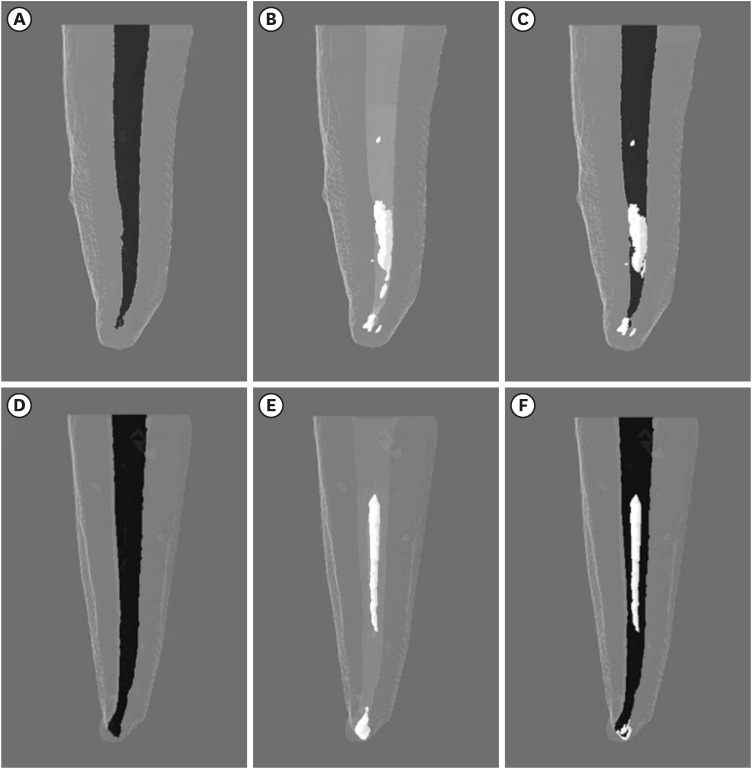
Figure 1 Three-dimensional pre- and post-endodontic retreatment images of representative specimens instrumented with reciprocating (A-C) and rotary (D-F) systems, illustrating the obturation (A, D) amount, spatial location of the remaining filling material (B, F), and superposition of the images (C, F).
Cited by 1 articles
-
Micro-CT evaluation of the removal of root fillings using rotary and reciprocating systems supplemented by XP-Endo Finisher, the Self-Adjusting File, or Er,Cr:YSGG laser
Gülsen Kiraz, Bulem Üreyen Kaya, Mert Ocak, Muhammet Bora Uzuner, Hakan Hamdi Çelik
Restor Dent Endod. 2023;48(4):e36. doi: 10.5395/rde.2023.48.e36.
Reference
-
1. Wilcox LR, Krell KV, Madison S, Rittman B. Endodontic retreatment: evaluation of gutta-percha and sealer removal and canal reinstrumentation. J Endod. 1987; 13:453–457. PMID: 3482104.
Article2. Stabholz A, Friedman S. Endodontic retreatment--case selection and technique. Part 2: treatment planning for retreatment. J Endod. 1988; 14:607–614. PMID: 3270681.
Article3. Friedman S, Stabholz A. Endodontic retreatment--case selection and technique. Part 1: criteria for case selection. J Endod. 1986; 12:28–33. PMID: 3456420.
Article4. Giuliani V, Cocchetti R, Pagavino G. Efficacy of ProTaper universal retreatment files in removing filling materials during root canal retreatment. J Endod. 2008; 34:1381–1384. PMID: 18928852.
Article5. Hammad M, Qualtrough A, Silikas N. Three-dimensional evaluation of effectiveness of hand and rotary instrumentation for retreatment of canals filled with different materials. J Endod. 2008; 34:1370–1373. PMID: 18928849.
Article6. Roggendorf MJ, Legner M, Ebert J, Fillery E, Frankenberger R, Friedman S. Micro-CT evaluation of residual material in canals filled with Activ GP or GuttaFlow following removal with NiTi instruments. Int Endod J. 2010; 43:200–209. PMID: 20158531.
Article7. Abramovitz I, Relles-Bonar S, Baransi B, Kfir A. The effectiveness of a self-adjusting file to remove residual gutta-percha after retreatment with rotary files. Int Endod J. 2012; 45:386–392. PMID: 22283664.
Article8. Rödig T, Hausdörfer T, Konietschke F, Dullin C, Hahn W, Hülsmann M. Efficacy of D-RaCe and ProTaper Universal Retreatment NiTi instruments and hand files in removing gutta-percha from curved root canals - a micro-computed tomography study. Int Endod J. 2012; 45:580–589. PMID: 22264204.
Article9. Solomonov M, Paqué F, Kaya S, Adigüzel O, Kfir A, Yiğit-Özer S. Self-adjusting files in retreatment: a high-resolution micro-computed tomography study. J Endod. 2012; 38:1283–1287. PMID: 22892752.
Article10. Cavenago BC, Ordinola-Zapata R, Duarte MA, del Carpio-Perochena AE, Villas-Bôas MH, Marciano MA, Bramante CM, Moraes IG. Efficacy of xylene and passive ultrasonic irrigation on remaining root filling material during retreatment of anatomically complex teeth. Int Endod J. 2014; 47:1078–1083. PMID: 24456216.
Article11. Fruchi LC, Ordinola-Zapata R, Cavenago BC, Hungaro Duarte MA, Bueno CE, De Martin AS. Efficacy of reciprocating instruments for removing filling material in curved canals obturated with a single-cone technique: a micro-computed tomographic analysis. J Endod. 2014; 40:1000–1004. PMID: 24935552.
Article12. Cardoso ER, Tookuni IV, Morais CA, Pavan NN, Santin GC, Capitanio M, Endo MS. Effectiveness of reciprocating and rotary retreatment files in the removal of endodontic filling material. Gen Dent. 2022; 70:22–25.13. Romeiro K, de Almeida A, Cassimiro M, Gominho L, Dantas E, Chagas N, Velozo C, Freire L, Albuquerque D. Reciproc and Reciproc Blue in the removal of bioceramic and resin-based sealers in retreatment procedures. Clin Oral Investig. 2020; 24:405–416.
Article14. Amoroso-Silva P, Alcalde MP, Hungaro Duarte MA, De-Deus G, Ordinola-Zapata R, Freire LG, Cavenago BC, De Moraes IG. Effect of finishing instrumentation using NiTi hand files on volume, surface area and uninstrumented surfaces in C-shaped root canal systems. Int Endod J. 2017; 50:604–611. PMID: 27194509.
Article15. Carr GB, Schwartz RS, Schaudinn C, Gorur A, Costerton JW. Ultrastructural examination of failed molar retreatment with secondary apical periodontitis: an examination of endodontic biofilms in an endodontic retreatment failure. J Endod. 2009; 35:1303–1309. PMID: 19720237.
Article16. Beasley RT, Williamson AE, Justman BC, Qian F. Time required to remove guttacore, thermafil plus, and thermoplasticized gutta-percha from moderately curved root canals with protaper files. J Endod. 2013; 39:125–128. PMID: 23228271.
Article17. Só MV, Saran C, Magro ML, Vier-Pelisser FV, Munhoz M. Efficacy of ProTaper retreatment system in root canals filled with gutta-percha and two endodontic sealers. J Endod. 2008; 34:1223–1225. PMID: 18793925.
Article18. Gergi R, Sabbagh C. Effectiveness of two nickel-titanium rotary instruments and a hand file for removing gutta-percha in severely curved root canals during retreatment: an ex vivo study. Int Endod J. 2007; 40:532–537. PMID: 17511787.
Article19. Barrieshi-Nusair KM. Gutta-percha retreatment: effectiveness of nickel-titanium rotary instruments versus stainless steel hand files. J Endod. 2002; 28:454–456. PMID: 12067128.
Article20. Zmener O, Pameijer CH, Banegas G. Retreatment efficacy of hand versus automated instrumentation in oval-shaped root canals: an ex vivo study. Int Endod J. 2006; 39:521–526. PMID: 16776756.
Article21. Schirrmeister JF, Wrbas KT, Schneider FH, Altenburger MJ, Hellwig E. Effectiveness of a hand file and three nickel-titanium rotary instruments for removing gutta-percha in curved root canals during retreatment. Oral Surg Oral Med Oral Pathol Oral Radiol Endod. 2006; 101:542–547. PMID: 16545721.
Article22. De-Deus G, Arruda TE, Souza EM, Neves A, Magalhães K, Thuanne E, Fidel RA. The ability of the Reciproc R25 instrument to reach the full root canal working length without a glide path. Int Endod J. 2013; 46:993–998. PMID: 23560929.
Article23. Crozeta BM, Silva-Sousa YT, Leoni GB, Mazzi-Chaves JF, Fantinato T, Baratto-Filho F, Sousa-Neto MD. Micro- computed tomography study of filling material removal from oval-shaped canals by using rotary, reciprocating, and adaptive motion systems. J Endod. 2016; 42:793–797. PMID: 26987688.
Article24. de Siqueira Zuolo A, Zuolo ML, da Silveira Bueno CE, Chu R, Cunha RS. Evaluation of the efficacy of TRUShape and Reciproc file systems in the removal of root filling material: an ex vivo micro-computed tomographic study. J Endod. 2016; 42:315–319. PMID: 26709199.
Article25. Özyürek T, Demiryürek EÖ. Efficacy of different nickel-titanium instruments in removing gutta-percha during root canal retreatment. J Endod. 2016; 42:646–649. PMID: 26898565.
Article26. Alves FR, Marceliano-Alves MF, Sousa JC, Silveira SB, Provenzano JC, Siqueira JF Jr. Removal of root canal fillings in curved canals using either reciprocating single- or rotary multi-instrument systems and a supplementary step with the XP-Endo Finisher. J Endod. 2016; 42:1114–1119. PMID: 27215810.
Article27. Vertucci FJ. Root canal anatomy of the human permanent teeth. Oral Surg Oral Med Oral Pathol. 1984; 58:589–599. PMID: 6595621.
Article
- Full Text Links
-
- Actions
-
Cited
- CITED
-
- Close
- Share
-
- Similar articles
-
- Effect of glide path preparation with PathFile and ProGlider on the cyclic fatigue resistance of WaveOne nickel-titanium files
- Assessment of postoperative pain after single-visit root canal treatment using rotary and reciprocating file systems: an in vivo study
- The effect of root canal preparation on the surface roughness of WaveOne and WaveOne Gold files: atomic force microscopy study
- Micro-CT evaluation of the removal of root fillings using rotary and reciprocating systems supplemented by XP-Endo Finisher, the Self-Adjusting File, or Er,Cr:YSGG laser
- Effectiveness and safety of rotary and reciprocating kinematics for retreatment of curved root canals: a systematic review of in vitro studies
