Leukocyte platelet-rich fibrin in endodontic microsurgery: a report of 2 cases
- Affiliations
-
- 1Department of Endodontics, Faculdade de Medicina Dentária, Universidade de Lisboa, Lisboa, Portugal
- 2Unidade de Investigação em Ciências Orais e Biomédicas (UICOB), Faculdade de Medicina Dentária, Universidade de Lisboa, Lisboa, Portugal
- KMID: 2548121
- DOI: http://doi.org/10.5395/rde.2022.47.e17
Abstract
- Endodontic microsurgery is a predictable treatment option when orthograde treatment or retreatment is unsuccessful or unfeasible. However, when there is a gross compromise of periapical bone, achievement of bone regeneration after the surgical procedure may be hampered. In such cases, the application of guided tissue regeneration principles, with adjunctive use of leukocyte platelet-rich fibrin to fill the bone defect as a bone substitute and as a membrane to cover the site, provides a cost-effective solution with the benefits of accelerated physiological healing and reduced post-surgical pain and discomfort. This case report presents 2 cases of endodontic microsurgery of the upper lateral incisors with loss of buccal cortical plate, where platelet-rich fibrin was successfully applied.
Figure
-
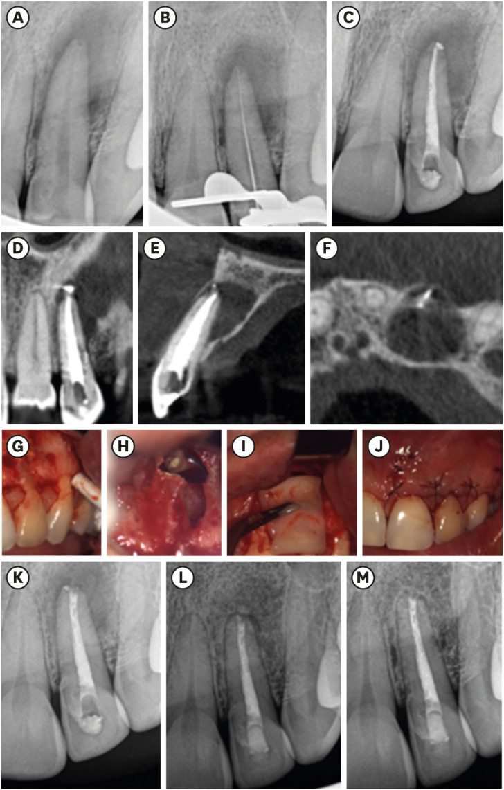
Figure 1 Chronological documentation of case 1: (A) initial periapical radiograph of tooth #22; (B) working length confirmation; (C) periapical radiograph showing the final root canal treatment; (D, E, F) axial, sagittal, and coronal views of cone-beam computed tomography prior to endodontic microsurgery; (G) mucoperiosteal flap; (H) apicoectomy, retrograde preparations, and obturation with MTA; (I) previously prepared leukocyte platelet-rich fibrin membranes used to fill and cover the surgical bone defect; (J) repositioned flap; (K) post-surgical periapical radiograph; (L) radiograph from a 6-month follow-up visit; (M) radiograph from a 12-month follow-up after endodontic microsurgery.
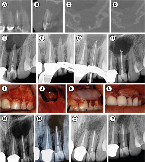
Figure 2 Chronological documentation of case 2: (A, B, C, D) Axial, sagittal, and coronal views of cone-beam computed tomography before root canal treatment; (E) initial periapical radiograph of tooth #22; (F) cleaned root canal after irrigation; (G) working length confirmation; (H) periapical radiograph showing the final root canal treatment; (I) mucoperiosteal flap; (J) apicoectomy, retrograde preparations, and obturation with mineral trioxide aggregate; (K) leukocyte platelet-rich fibrin membranes filling the bone defect; (L) repositioning of the surgical flap; (M) radiograph from a 2-month follow-up; (N) radiograph from a 4-month follow-up; (O) radiograph from a 6-month follow-up; (P) and radiograph from a 12-month follow-up after endodontic microsurgery.
Reference
-
1. Haapasalo M, Endal U, Zandi H, Coil JM. Eradication of endodontic infection by instrumentation and irrigation solutions. Endod Topics. 2005; 10:77–102.2. Siqueira JF Jr, Rôças IN. Clinical implications and microbiology of bacterial persistence after treatment procedures. J Endod. 2008; 34:1291–1301.e3. PMID: 18928835.
Article3. Schilder H. Filling root canals in three dimensions. 1967. J Endod. 2006; 32:281–290. PMID: 16554195.4. Nair PN. On the causes of persistent apical periodontitis: a review. Int Endod J. 2006; 39:249–281. PMID: 16584489.
Article5. Kang M, In Jung H, Song M, Kim SY, Kim HC, Kim E. Outcome of nonsurgical retreatment and endodontic microsurgery: a meta-analysis. Clin Oral Investig. 2015; 19:569–582.6. Tsesis I, Rosen E, Taschieri S, Telishevsky Strauss Y, Ceresoli V, Del Fabbro M. Outcomes of surgical endodontic treatment performed by a modern technique: an updated meta-analysis of the literature. J Endod. 2013; 39:332–339. PMID: 23402503.
Article7. European Society of Endodontology. Quality guidelines for endodontic treatment: consensus report of the European Society of Endodontology. Int Endod J. 2006; 39:921–930. PMID: 17180780.8. Song M, Kim SG, Shin SJ, Kim HC, Kim E. The influence of bone tissue deficiency on the outcome of endodontic microsurgery: a prospective study. J Endod. 2013; 39:1341–1345. PMID: 24139252.
Article9. von Arx T, Cochran DL. Rationale for the application of the GTR principle using a barrier membrane in endodontic surgery: a proposal of classification and literature review. Int J Periodontics Restorative Dent. 2001; 21:127–139. PMID: 11829387.10. Del Fabbro M, Corbella S, Sequeira-Byron P, Tsesis I, Rosen E, Lolato A, Taschieri S. Endodontic procedures for retreatment of periapical lesions. Cochrane Database Syst Rev. 2016; 10:CD005511. PMID: 27759881.11. Karan NB, Aricioğlu B. Assessment of bone healing after mineral trioxide aggregate and platelet-rich fibrin application in periapical lesions using cone-beam computed tomographic imaging. Clin Oral Investig. 2020; 24:1065–1072.
Article12. Meschi N, Castro AB, Vandamme K, Quirynen M, Lambrechts P. The impact of autologous platelet concentrates on endodontic healing: a systematic review. Platelets. 2016; 27:613–633. PMID: 27658056.
Article13. Del Fabbro M, Ceresoli V, Lolato A, Taschieri S. Effect of platelet concentrate on quality of life after periradicular surgery: a randomized clinical study. J Endod. 2012; 38:733–739. PMID: 22595104.
Article14. Song M, Kim HC, Lee W, Kim E. Analysis of the cause of failure in nonsurgical endodontic treatment by microscopic inspection during endodontic microsurgery. J Endod. 2011; 37:1516–1519. PMID: 22000454.
Article15. de Chevigny C, Dao TT, Basrani BR, Marquis V, Farzaneh M, Abitbol S, Friedman S. Treatment outcome in endodontics: the Toronto study--phases 3 and 4: orthograde retreatment. J Endod. 2008; 34:131–137. PMID: 18215667.
Article16. Huang S, Chen NN, Yu VS, Lim HA, Lui JN. Long-term success and survival of endodontic microsurgery. J Endod. 2020; 46:149–157.e4. PMID: 31879031.
Article17. Pecora G, Baek SH, Rethnam S, Kim S. Barrier membrane techniques in endodontic microsurgery. Dent Clin North Am. 1997; 41:585–602. PMID: 9248693.
Article18. von Arx T, Hänni S, Jensen SS. Correlation of bone defect dimensions with healing outcome one year after apical surgery. J Endod. 2007; 33:1044–1048. PMID: 17931929.
Article19. Choukroun J, Diss A, Simonpieri A, Girard MO, Schoeffler C, Dohan SL, Dohan AJ, Mouhyi J, Dohan DM. Platelet-rich fibrin (PRF): a second-generation platelet concentrate. Part IV: clinical effects on tissue healing. Oral Surg Oral Med Oral Pathol Oral Radiol Endod. 2006; 101:e56–e60. PMID: 16504852.
Article20. Molven O, Halse A, Grung B. Incomplete healing (scar tissue) after periapical surgery--radiographic findings 8 to 12 years after treatment. J Endod. 1996; 22:264–268. PMID: 8632141.
Article
- Full Text Links
-
- Actions
-
Cited
- CITED
-
- Close
- Share
-
- Similar articles
-
- Platelet rich fibrin - a novel acumen into regenerative endodontic therapy
- Clinical effectiveness of combining platelet rich fibrin with alloplastic bone substitute for the management of combined endodontic periodontal lesion
- Evaluation of blood clot, plateletrich plasma, and platelet-rich fibrin– mediated regenerative endodontic procedures in teeth with periapical pathology: a CBCT study
- Effect of Platelet-rich Plasma on Burn Wounds according to Time of Application: An Experimental Study on Rats
- Fibrin Clot Delivery System for Meniscal Repair
