Restor Dent Endod.
2022 Feb;47(1):e8. 10.5395/rde.2022.47.e8.
Comparison of instrumental methods for color change assessment of Giomer resins
- Affiliations
-
- 1Post-Graduate Program, School of Dentistry, Universidade Federal de Minas Gerais, Belo Horizonte, Brazil
- 2Department of Restorative Dentistry, School of Dentistry, Universidade Federal de Minas Gerais, Belo Horizonte, Brazil
- 3Department of Prosthodontics, School of Dentistry, Centro Universitário de Belo Horizonte, Belo Horizonte, Brazil
- 4Department of Restorative Dentistry, Faculty of Dental Medicine, Hokkaido University, Sapporo, Japan
- 5Department of Clinic, Pathology and Dental Surgery, School of Dentistry, Universidade Federal de Minas Gerais, Belo Horizonte, Brazil
- KMID: 2548117
- DOI: http://doi.org/10.5395/rde.2022.47.e8
Abstract
Objectives
The aim of this study was to compare the color change of the Giomer resin composite (Beautifil-Bulk) by using photographs obtained with a smartphone (iPhone 6S) associated with Adobe Photoshop software (digital method), with the spectrophotometric method (Vita Easyshade) after immersion in different pigment solutions.
Materials and Methods
Twenty resin composite samples with a diameter of 15.0 mm and thickness of 1.0 mm were confectioned in A2 color (n = 5). Photographs and initial color readings were performed with a smartphone and spectrophotometer, respectively. Then, samples were randomly divided and subjected to cycles of immersion in distilled water (control), açai, Coke, and tomato sauce, 3 times a day, 20 minutes for 7 days. Later, new photographs and color readings were taken.
Results
The analysis (2-way analysis of variance, Holm-Sidak, p < 0.05) demonstrated no statistical difference (p < 0.005) between the methods in all groups. Similar color changes were observed for all pigment solutions when using the spectrophotometric method. For the digital method, all color changes were clinically unacceptable, with distilled water and tomato sauce similar to each other and with statistical differences (p < 0.005) for Coke and açai.
Conclusions
Only the tomato sauce produced a color change above the acceptability threshold using both methods of color assessment. The spectrophotometric and digital methods produce different patterns of color change. According to our results, the spectrophotometric method is more recommended in color change assessment.
Keyword
Figure
-
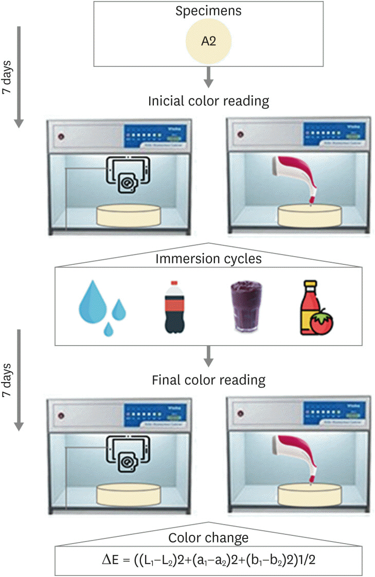
Figure 1 Flowchart summarizing the methodology.
Reference
-
1. Paravina RD, Ghinea R, Herrera LJ, Bona AD, Igiel C, Linninger M, Sakai M, Takahashi H, Tashkandi E, Perez MM. Color difference thresholds in dentistry. J Esthet Restor Dent. 2015; 27(Supplement 1):S1–S9. PMID: 25886208.
Article2. Chen H, Huang J, Dong X, Qian J, He J, Qu X, Lu E. A systematic review of visual and instrumental measurements for tooth shade matching. Quintessence Int. 2012; 43:649–659. PMID: 23034418.3. McLaren EA, Figueira J, Goldstein RE. A technique using calibrated photography and photoshop for accurate shade analysis and communication. Compend Contin Educ Dent. 2017; 38:106–113. PMID: 28156124.4. Brandt J, Nelson S, Lauer HC, von Hehn U, Brandt S. In vivo study for tooth colour determination-visual versus digital. Clin Oral Investig. 2017; 21:2863–2871.5. Perroni AP, Bergoli CD, Dos Santos MB, Moraes RR, Boscato N. Spectrophotometric analysis of clinical factors related to the color of ceramic restorations: a pilot study. J Prosthet Dent. 2017; 118:611–616. PMID: 28385444.
Article6. Miyajiwala JS, Kheur MG, Patankar AH, Lakha TA. Comparison of photographic and conventional methods for tooth shade selection: a clinical evaluation. J Indian Prosthodont Soc. 2017; 17:273–281. PMID: 28936042.
Article7. Alsaleh S, Labban M, AlHariri M, Tashkandi E. Evaluation of self shade matching ability of dental students using visual and instrumental means. J Dent. 2012; 40(Supplement 1):e82–e87. PMID: 22306532.
Article8. de Bragança RM, Moraes RR, Faria-E-Silva AL. Color assessment of resin composite by using cellphone images compared with a spectrophotometer. Restor Dent Endod. 2021; 46:e23. PMID: 34123759.
Article9. International Organization for Standardization (ISO). ISO 4049:2019. Dentistry — Polymer-based restorative materials. 3rd ed. Geneve: ISO;2000.10. Lee YK, Yu B, Lee SH, Cho MS, Lee CY, Lim HN. Shade compatibility of esthetic restorative materials--a review. Dent Mater. 2010; 26:1119–1126. PMID: 20832851.
Article11. Choi JW, Lee MJ, Oh SH, Kim KM. Changes in the physical properties and color stability of aesthetic restorative materials caused by various beverages. Dent Mater J. 2019; 38:33–40. PMID: 30298856.
Article12. Tekçe N, Tuncer S, Demirci M, Serim ME, Baydemir C. The effect of different drinks on the color stability of different restorative materials after one month. Restor Dent Endod. 2015; 40:255–261. PMID: 26587410.
Article13. Commission Internationale de l’Eclairage (CIE). CIE recommendations on uniform color spaces, color difference equations, psychometric color terms. 1st ed. Paris: CIE;1978.14. Sluzker A, Knösel M, Athanasiou AE. Sensitivity of digital dental photo CIE L*a*b* analysis compared to spectrophotometer clinical assessments over 6 months. Am J Dent. 2011; 24:300–304. PMID: 22165458.15. Chu SJ, Trushkowsky RD, Paravina RD. Dental color matching instruments and systems. Review of clinical and research aspects. J Dent. 2010; 38(Supplement 2):e2–ee16. PMID: 20621154.
Article16. Dozić A, Kleverlaan CJ, El-Zohairy A, Feilzer AJ, Khashayar G. Performance of five commercially available tooth color-measuring devices. J Prosthodont. 2007; 16:93–100. PMID: 17362418.
Article17. Kalantari MH, Ghoraishian SA, Mohaghegh M. Evaluation of accuracy of shade selection using two spectrophotometer systems: Vita Easyshade and Degudent Shadepilot. Eur J Dent. 2017; 11:196–200. PMID: 28729792.
Article18. Lehmann K, Devigus A, Wentaschek S, Igiel C, Scheller H, Paravina R. Comparison of visual shade matching and electronic color measurement device. Int J Esthet Dent. 2017; 12:396–404. PMID: 28717795.19. Anand D, Surendra Kumar GP, Anand DY, Sundar MK, Sharma R, Gaurav A. Shade selection: spectrophotometer vs digital camera – a comparative in-vitro study. IP Ann Prosthodont Restor Dent. 2016; 2:73–78.20. Tam WK, Lee HJ. Accurate shade image matching by using a smartphone camera. J Prosthodont Res. 2017; 61:168–176. PMID: 27553123.
Article21. Igiel C, Weyhrauch M, Wentaschek S, Scheller H, Lehmann KM. Dental color matching: a comparison between visual and instrumental methods. Dent Mater J. 2016; 35:63–69. PMID: 26830824.
Article22. Gonulol N, Ozer S, Sen Tunc E. Water sorption, solubility, and color stability of giomer restoratives. J Esthet Restor Dent. 2015; 27:300–306. PMID: 25145876.
Article23. Adusumilli H, Avula JS, Kakarla P, Bandi S, Mallela GM, Vallabhaneni K. Color stability of esthetic restorative materials used in pediatric dentistry: an in vitro study. J Indian Soc Pedod Prev Dent. 2016; 34:233–237. PMID: 27461806.
Article24. El-Sharkawy FM, Zaghlou NM, Ell-kappaney AM. Effect of water absorption on color stability of different resin based restorative materials in vitro study. Int J Compos Mater. 2012; 2:7–10.25. Moon JD, Seon EM, Son SA, Jung KH, Kwon YH, Park JK. Effect of immersion into solutions at various pH on the color stability of composite resins with different shades. Restor Dent Endod. 2015; 40:270–276. PMID: 26587412.
Article26. Soares-Geraldo D, Scaramucci T, Steagall W Jr, Braga SR, Sobral MA. Interaction between staining and degradation of a composite resin in contact with colored foods. Braz Oral Res. 2011; 25:369–375. PMID: 21860925.
Article27. Tonetto MR, Neto CS, Felício CM, Domingos PA, Campos EA, Andrade MF. Effect of staining agents on color change of composites. RSBO. 2012; 9:266–271.
- Full Text Links
-
- Actions
-
Cited
- CITED
-
- Close
- Share
-
- Similar articles
-
- Color stability of esthetic restorative materials after application of fluoride varnishes
- Color Comparison of Maxillary Primary Anterior Teeth and Various Composite Resins using a Spectrophotometer
- A study on the color stability of porcelain repair resins
- Understanding of the color in composite resin
- Evaluation of the color stability of light cured composite resins according to the resin matrices
