Effect of hydrogel-based antibiotic intracanal medicaments on crown discoloration
- Affiliations
-
- 1Department of Restorative Dentistry, College of Dental Medicine, Umm Al-Qura University, Makkah, Saudi Arabia
- 2Department of Biomedical Sciences and Comprehensive Care, Indiana University School of Dentistry, Indianapolis, IN, USA
- 3Department of Endodontics, Indiana University School of Dentistry, Indianapolis, IN, USA
- 4Department of Endodontics, Case School of Dental Medicine, Cleveland, OH, USA
- KMID: 2548096
- DOI: http://doi.org/10.5395/rde.2021.46.e52
Abstract
Objectives
This study evaluated the effects of low and moderate concentrations of triple antibiotic paste (TAP) and double antibiotic paste (DAP) loaded into a hydrogel system on crown discoloration and explored whether application of an adhesive bonding agent prevented crown discoloration.
Materials and Methods
Intact human molars (n = 160) were horizontally sectioned 1 mm apical to the cementoenamel junction. The crowns were randomized into 8 experimental groups (calcium hydroxide, Ca[OH]2 ; 1, 10, and 1,000 mg/mL TAP and DAP; and no medicament. The pulp chambers in half of the samples were coated with an adhesive bonding agent before receiving the intracanal medicament. Color changes (ΔE) were detected by spectrophotometry after 1 day, 1 week, and 4 weeks, and after 5,000 thermal cycles, with ΔE = 3.7 as a perceptible threshold. The 1-sample t-test was used to determine the significance of color changes relative to 3.7. Analysis of variance was used to evaluate the effects of treatment, adhesive, and time on color change, and the level of significance was p < 0.05.
Results
Ca(OH)2 and 1 and 10 mg/mL DAP did not cause clinically perceivable tooth discoloration. Adhesive agent use significantly decreased tooth discoloration in the 1,000 mg/mL TAP group up to 4 weeks. However, adhesive use did not significantly improve coronal discoloration after thermocycling when 1,000 mg/mL TAP was used.
Conclusions
Ca(OH)2 and 1 and 10 mg/mL DAP showed no clinical discoloration. Using an adhesive significantly improved coronal discoloration up to 4 weeks with 1,000 mg/mL TAP.
Figure
-
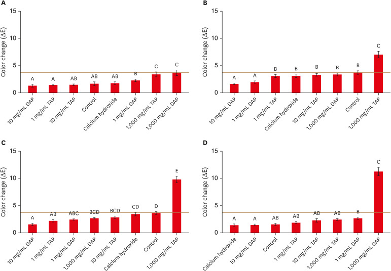
Figure 1 Mean ± standard error of color changes in different treatment groups with adhesive after intracanal medicament application for (A) 1 day, (B) 1 week, (C) 4 weeks, and (D) after thermal cycling. Treatments with the same superscript letter were not significantly different at p < 0.05. The red horizontal line represents the perceptible threshold (ΔE = 3.7).
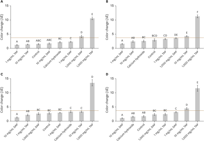
Figure 2 Mean ± standard error of color changes in different treatment groups without adhesive after intracanal medicament application for (A) 1 day, (B) 1 week, (C) 4 weeks, and (D) after thermal cycling. Treatments with the same superscript letter were not significantly different at p < 0.05. The red horizontal line represents the perceptible threshold (ΔE = 3.7).
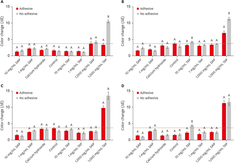
Figure 3 Mean ± standard error of color changes in the adhesive and no adhesive treatment groups after intracanal medicament application for (A) 1 day, (B) 1 week, (C) 4 weeks, and (D) after thermal cycling. The adhesive and no adhesive treatment groups with the same superscript letter were not significantly different at p < 0.05. The red horizontal line represents the perceptible threshold (ΔE = 3.7).
Reference
-
1. Thibodeau B, Trope M. Pulp revascularization of a necrotic infected immature permanent tooth: case report and review of the literature. Pediatr Dent. 2007; 29:47–50. PMID: 18041512.2. Lenzi R, Trope M. Revitalization procedures in two traumatized incisors with different biological outcomes. J Endod. 2012; 38:411–414. PMID: 22341086.
Article3. Ding RY, Cheung GS, Chen J, Yin XZ, Wang QQ, Zhang CF. Pulp revascularization of immature teeth with apical periodontitis: a clinical study. J Endod. 2009; 35:745–749. PMID: 19410097.
Article4. Fouad AF, Verma P. Healing after regenerative procedures with and without pulpal infection. J Endod. 2014; 40:S58–S64. PMID: 24698695.
Article5. Wigler R, Kaufman AY, Lin S, Steinbock N, Hazan-Molina H, Torneck CD. Revascularization: a treatment for permanent teeth with necrotic pulp and incomplete root development. J Endod. 2013; 39:319–326. PMID: 23402501.
Article6. Law AS. Considerations for regeneration procedures. J Endod. 2013; 39:S44–S56. PMID: 23439044.
Article7. Chen MY, Chen KL, Chen CA, Tayebaty F, Rosenberg PA, Lin LM. Responses of immature permanent teeth with infected necrotic pulp tissue and apical periodontitis/abscess to revascularization procedures. Int Endod J. 2012; 45:294–305. PMID: 22077958.
Article8. Lovelace TW, Henry MA, Hargreaves KM, Diogenes A. Evaluation of the delivery of mesenchymal stem cells into the root canal space of necrotic immature teeth after clinical regenerative endodontic procedure. J Endod. 2011; 37:133–138. PMID: 21238791.
Article9. Miller EK, Lee JY, Tawil PZ, Teixeira FB, Vann WF Jr. Emerging therapies for the management of traumatized immature permanent incisors. Pediatr Dent. 2012; 34:66–69. PMID: 22353461.10. Trope M. Treatment of the immature tooth with a non-vital pulp and apical periodontitis. Dent Clin North Am. 2010; 54:313–324. PMID: 20433980.
Article11. Kohli MR, Yamaguchi M, Setzer FC, Karabucak B. Spectrophotometric analysis of coronal tooth discoloration induced by various bioceramic cements and other endodontic materials. J Endod. 2015; 41:1862–1866. PMID: 26386949.
Article12. Lenherr P, Allgayer N, Weiger R, Filippi A, Attin T, Krastl G. Tooth discoloration induced by endodontic materials: a laboratory study. Int Endod J. 2012; 45:942–949. PMID: 22506849.
Article13. Kim JH, Kim Y, Shin SJ, Park JW, Jung IY. Tooth discoloration of immature permanent incisor associated with triple antibiotic therapy: a case report. J Endod. 2010; 36:1086–1091. PMID: 20478471.
Article14. Akcay M, Arslan H, Yasa B, Kavrık F, Yasa E. Spectrophotometric analysis of crown discoloration induced by various antibiotic pastes used in revascularization. J Endod. 2014; 40:845–848. PMID: 24862714.
Article15. Reynolds K, Johnson JD, Cohenca N. Pulp revascularization of necrotic bilateral bicuspids using a modified novel technique to eliminate potential coronal discolouration: a case report. Int Endod J. 2009; 42:84–92. PMID: 19125982.
Article16. Poly A, Marques F, Fidel SR, Monnerat AF, Sassone LM. Ability of two single-step restorative materials to avoid crown darkening caused by intracanal minocycline paste. Clin Oral Investig. 2019; 23:1281–1286.
Article17. Afkhami F, Elahy S, Nahavandi AM, Kharazifard MJ, Sooratgar A. Discoloration of teeth due to different intracanal medicaments. Restor Dent Endod. 2019; 44:e10. PMID: 30834232.
Article18. Dettwiler CA, Walter M, Zaugg LK, Lenherr P, Weiger R, Krastl G. In vitro assessment of the tooth staining potential of endodontic materials in a bovine tooth model. Dent Traumatol. 2016; 32:480–487. PMID: 27188429.
Article19. Kim ST, Abbott PV, McGinley P. The effects of Ledermix paste on discolouration of immature teeth. Int Endod J. 2000; 33:233–237. PMID: 11307440.
Article20. Esmaeili B, Alaghehmand H, Kordafshari T, Daryakenari G, Ehsani M, Bijani A. Coronal discoloration induced by calcium-enriched mixture, mineral trioxide aggregate and calcium hydroxide: a spectrophotometric analysis. Iran Endod J. 2016; 11:23–28. PMID: 26843873.21. Tanase S, Tsuchiya H, Yao J, Ohmoto S, Takagi N, Yoshida S. Reversed-phase ion-pair chromatographic analysis of tetracycline antibiotics. Application to discolored teeth. J Chromatogr B Biomed Sci Appl. 1998; 706:279–285. PMID: 9551814.
Article22. AlSaeed T, Nosrat A, Melo MA, Wang P, Romberg E, Xu H, Fouad AF. Antibacterial efficacy and discoloration potential of endodontic topical antibiotics. J Endod. 2018; 44:1110–1114. PMID: 29803336.
Article23. Shokouhinejad N, Razmi H, Farbod M, Alikhasi M, Camilleri J. Coronal tooth discoloration induced by regenerative endodontic treatment using different scaffolds and intracanal coronal barriers: a 6-month ex vivo study. Restor Dent Endod. 2019; 44:e25. PMID: 31485421.
Article24. Shokouhinejad N, Khoshkhounejad M, Alikhasi M, Bagheri P, Camilleri J. Prevention of coronal discoloration induced by regenerative endodontic treatment in an ex vivo model. Clin Oral Investig. 2018; 22:1725–1731.
Article25. Alghilan MA, Windsor LJ, Palasuk J, Yassen GH. Attachment and proliferation of dental pulp stem cells on dentine treated with different regenerative endodontic protocols. Int Endod J. 2017; 50:667–675. PMID: 27272393.
Article26. Ruparel NB, Teixeira FB, Ferraz CC, Diogenes A. Direct effect of intracanal medicaments on survival of stem cells of the apical papilla. J Endod. 2012; 38:1372–1375. PMID: 22980180.
Article27. Yassen GH, Eckert GJ, Platt JA. Effect of intracanal medicaments used in endodontic regeneration procedures on microhardness and chemical structure of dentin. Restor Dent Endod. 2015; 40:104–112. PMID: 25984471.
Article28. Prather BT, Ehrlich Y, Spolnik K, Platt JA, Yassen GH. Effects of two combinations of triple antibiotic paste used in endodontic regeneration on root microhardness and chemical structure of radicular dentine. J Oral Sci. 2014; 56:245–251. PMID: 25500921.
Article29. Humel MMC, Oliveira MT, Cavalli V, Giannini M. Effect of storage and disinfection methods of extracted bovine teeth on bond strength to dentin. Braz J Oral Sci. 2016; 6:1402–1406.30. Jarahi N, Borouziniat A, Jarahi L, Nejat AH. Effect of different storage solutions and autoclaving on shear bond strength of composite to dentin. J Res Med Dent Sci. 2018; 6:50–53.31. Gale MS, Darvell BW. Thermal cycling procedures for laboratory testing of dental restorations. J Dent. 1999; 27:89–99. PMID: 10071465.32. International Organization for Standardization. ISO/TR 11405. Dental materials-guidance on testing of adhesive to tooth structure. Geneva: International Organization for Standardization;1994. p. 1–14.33. Johnston WM, Kao EC. Assessment of appearance match by visual observation and clinical colorimetry. J Dent Res. 1989; 68:819–822. PMID: 2715476.
Article34. Fundaoğlu Küçükekenci F, Küçükekenci AS, Çakici F. Evaluation of the preventive efficacy of three dentin tubule occlusion methods against discoloration caused by triple-antibiotic paste. Odontology. 2019; 107:186–189. PMID: 30171400.35. Santos LG, Felippe WT, Souza BD, Konrath AC, Cordeiro MM, Felippe MC. Crown discoloration promoted by materials used in regenerative endodontic procedures and effect of dental bleaching: spectrophotometric analysis. J Appl Oral Sci. 2017; 25:234–242. PMID: 28403365.36. Fundaoğlu Küçükekenci F, Çakici F, Küçükekenci AS. Spectrophotometric analysis of discoloration and internal bleaching after use of different antibiotic pastes. Clin Oral Investig. 2019; 23:161–167.37. Cho WC, Kim MS, Lee HS, Choi SC, Nam OH. Pulp revascularization of a severely malformed immature maxillary canine. J Oral Sci. 2016; 58:295–298. PMID: 27349553.
Article38. Tekce N, Demirci M, Tuncer S, Uysal Ö. Microtensile bond strength and sealing efficiency of all-in-one self-etching adhesives. Biotechnol Biotechnol Equip. 2015; 29:570–578.
Article39. Poggio C, Beltrami R, Scribante A, Colombo M, Chiesa M. Shear bond strength of one-step self-etch adhesives: pH influence. Dent Res J (Isfahan). 2015; 12:209–214. PMID: 26005459.
- Full Text Links
-
- Actions
-
Cited
- CITED
-
- Close
- Share
-
- Similar articles
-
- Discoloration of teeth due to different intracanal medicaments
- Effect of Intracanal Medicaments on Push-out Bond Strength of Calcium Silicate-based Materials
- Effect of intracanal medicaments used in endodontic regeneration procedures on microhardness and chemical structure of dentin
- Prevention of tooth discoloration associated with triple antibiotics
- Coronal tooth discoloration induced by regenerative endodontic treatment using different scaffolds and intracanal coronal barriers: a 6-month ex vivo study
