Errors in light-emitting diodes positioning when curing bulk fill and incremental composites: impact on properties after aging
- Affiliations
-
- 1Ph.D Program Dental Biomedical Sciences, University of Maryland School of Dentistry, Baltimore, MD, USA
- 2Division of Operative Dentistry, Department of General Dentistry, University of Maryland School of Dentistry, Baltimore, MD, USA
- 3Advanced Education in General Dentistry Division, Department of General Dentistry, University of Maryland School of Dentistry, Baltimore, MD, USA
- 4Department of Dental Materials, School of Dentistry, Federal University of Rio Grande do Sul, Porto Alegre, RS, Brazil
- KMID: 2548095
- DOI: http://doi.org/10.5395/rde.2021.46.e51
Abstract
Objectives
This study aimed to evaluate the effect of improper positioning single-peak and multi-peak lights on color change, microhardness of bottom and top, and surface topography of bulk fill and incremental composites after artificial aging for 1 year.
Materials and Methods
Bulk fill and incremental composites were cured using multi-peak and single-peak light-emitting diode (LED) following 4 clinical conditions: (1) optimal condition (no angulation or tip displacement), (2) tip-displacement (2 mm), (3) slight tip angulation (α = 20°) and (4) moderate tip angulation (α = 35°). After 1-year of water aging, the specimens were analyzed for color changes (ΔE), Vickers hardness, surface topography (Ra, Rt, and Rv), and scanning electron microscopy.
Results
For samples cured by single-peak LED, the improper positioning significantly increases the color change compared to the optimal position regardless of the type of composite (p < 0.001). For multi-peak LED, the type of resin composite and the curing condition displayed a significant effect on ΔE (p < 0.001). For both LEDs, the Vickers hardness and bottom/top ratio of Vickers hardness were affected by the type of composite and the curing condition (p < 0.01).
Conclusions
The bulk fill composite presented greater resistance to wear, higher color stability, and better microhardness than the incremental composite when subjected to improper curing. The multi-peak LED improves curing under improper conditions compared to single-peak LED. Prevention of errors when curing composites requires the attention of all personnel involved in the patient's care once the clinical relevance of the appropriate polymerization reflects on reliable long-term outcomes.
Keyword
Figure
-
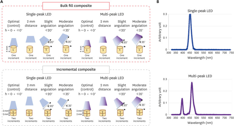
Figure 1 Schematic drawing showing the design of the study. Bulk fill and incremental composites were cured with single-peak and multi-peak LCUs following different conditions (optimal, 2 mm distance, slight angulation, and moderate angulation). The samples were prepared using customized 3D molds to standardization of the distance and angulations. The design of each mold was elaborated to allow the LCU tip to maintain the distance or the specific angulation (20° or 35°) in relation to the composite surface. The composites were aged for 1 year, and then the color stability, Vickers hardness, bottom/top ratio of Vickers hardness, and surface topography parameters were investigated.LED, light-emitting diode; LCU, light-curing unit.
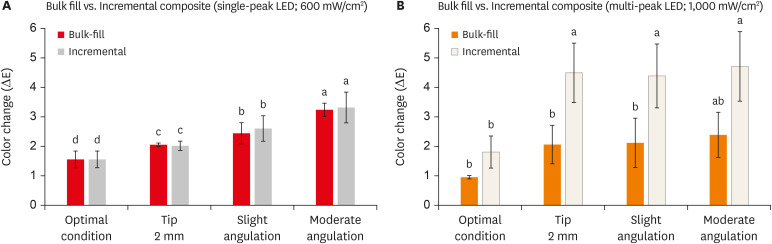
Figure 2 The color change is expressed by ΔE of the bulk fill and incremental composites cured using single-peak (A) and multi-peak (B) following different curing conditions. Values indicated by different letters are statistically different from each other (p < 0.05).LED, light-emitting diode.
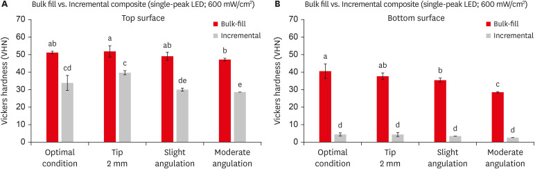
Figure 3 The vickers hardness values of the top (A) and bottom (B) surfaces of bulk fill and incremental composites cured using single-peak light-curing unit following different curing conditions. Values indicated by different letters are statistically different from each other (p < 0.05).LED, light-emitting diode.
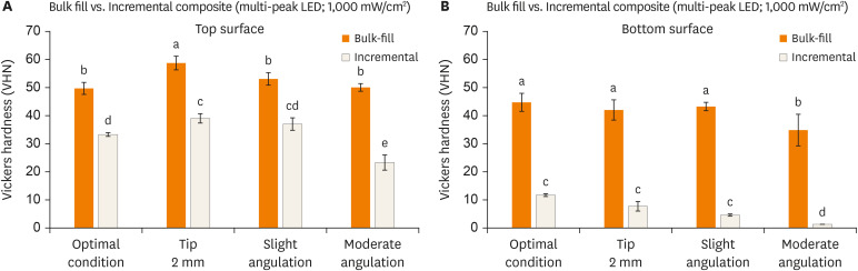
Figure 4 The Vickers hardness values of the top (A) and bottom (B) surfaces of bulk fill and incremental composites cured using multi-peak light-curing unit following different curing conditions. Values indicated by different letters are statistically different from each other (p < 0.05).LED, light-emitting diode.
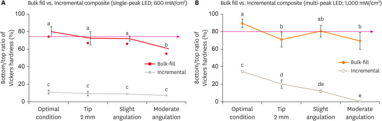
Figure 5 The bottom/top ratio of Vickers hardness of bulk fill and incremental composite cured using single-peak (A) and multi-peak (B) following different curing conditions. Values indicated by different letters are statistically different from each other (p < 0.05).LED, light-emitting diode.
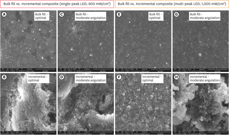
Figure 6 Scanning electron microscopy images for bulk fill and incremental composites cured with single-peak (A-D) and multi-peak (E-H) light-curing unit following the optimal and moderate angulation conditions. The incremental composite was associated with excessive topography changes and alterations compared to bulk fill composites.LED, light-emitting diode.
Reference
-
1. AlShaafi MM. Factors affecting polymerization of resin-based composites: a literature review. Saudi Dent J. 2017; 29:48–58. PMID: 28490843.
Article2. Price RB, Ferracane JL, Shortall AC. Light-curing units: a review of what we need to know. J Dent Res. 2015; 94:1179–1186. PMID: 26156516.3. de Oliveira DC, de Menezes LR, Gatti A, Correr Sobrinho L, Ferracane JL, Sinhoreti MA. Effect of nanofiller loading on cure efficiency and potential color change of model composites. J Esthet Restor Dent. 2016; 28:171–177. PMID: 26880567.
Article4. Alrahlah A, Silikas N, Watts DC. Post-cure depth of cure of bulk fill dental resin-composites. Dent Mater. 2014; 30:149–154. PMID: 24268044.
Article5. Gan JK, Yap AU, Cheong JW, Arista N, Tan C. Bulk-fill composites: effectiveness of cure with poly- and monowave curing lights and modes. Oper Dent. 2018; 43:136–143. PMID: 28976838.
Article6. Aggarwal N, Jain A, Gupta H, Abrol A, Singh C, Rapgay T. The comparative evaluation of depth of cure of bulk-fill composites - an in vitro study. J Conserv Dent. 2019; 22:371–375. PMID: 31802822.
Article7. Van Ende A, De Munck J, Van Landuyt KL, Poitevin A, Peumans M, Van Meerbeek B. Bulk-filling of high C-factor posterior cavities: effect on adhesion to cavity-bottom dentin. Dent Mater. 2013; 29:269–277. PMID: 23228335.
Article8. Flury S, Hayoz S, Peutzfeldt A, Hüsler J, Lussi A. Depth of cure of resin composites: is the ISO 4049 method suitable for bulk fill materials? Dent Mater. 2012; 28:521–528. PMID: 22391146.
Article9. Azzopardi N, Moharamzadeh K, Wood DJ, Martin N, van Noort R. Effect of resin matrix composition on the translucency of experimental dental composite resins. Dent Mater. 2009; 25:1564–1568. PMID: 19709725.
Article10. Price RB, Ferracane JL, Hickel R, Sullivan B. The light-curing unit: an essential piece of dental equipment. Int Dent J. 2020; 70:407–417. PMID: 32696512.
Article11. Derchi G, Vano M, Ceseracciu L, Diaspro A, Salerno M. Stiffness effect of using polywave or monowave LED units for photo-curing different bulk fill composites. Dent Mater J. 2018; 37:709–716. PMID: 30047507.
Article12. Menees TS, Lin CP, Kojic DD, Burgess JO, Lawson NC. Depth of cure of bulk fill composites with monowave and polywave curing lights. Am J Dent. 2015; 28:357–361. PMID: 26897758.13. Maktabi H, Balhaddad AA, Alkhubaizi Q, Strassler H, Melo MAS. Factors influencing success of radiant exposure in light-curing posterior dental composite in the clinical setting. Am J Dent. 2018; 31:320–328. PMID: 30658380.14. Konerding KL, Heyder M, Kranz S, Guellmar A, Voelpel A, Watts DC, Jandt KD, Sigusch BW. Study of energy transfer by different light curing units into a class III restoration as a function of tilt angle and distance, using a MARC Patient Simulator (PS). Dent Mater. 2016; 32:676–686. PMID: 27017156.
Article15. Al-Zain AO, Eckert GJ, Lukic H, Megremis S, Platt JA. Polymerization pattern characterization within a resin-based composite cured using different curing units at two distances. Clin Oral Investig. 2019; 23:3995–4010.
Article16. Eshmawi YT, Al-Zain AO, Eckert GJ, Platt JA. Variation in composite degree of conversion and microflexural strength for different curing lights and surface locations. J Am Dent Assoc. 2018; 149:893–902. PMID: 30149887.
Article17. Janda R, Roulet JF, Latta M, Steffin G, Rüttermann S. Color stability of resin-based filling materials after aging when cured with plasma or halogen light. Eur J Oral Sci. 2005; 113:251–257. PMID: 15953251.
Article18. Maktabi H, Ibrahim M, Alkhubaizi Q, Weir M, Xu H, Strassler H, Fugolin APP, Pfeifer CS, Melo MAS. Underperforming light curing procedures trigger detrimental irradiance-dependent biofilm response on incrementally placed dental composites. J Dent. 2019; 88:103110. PMID: 31022421.
Article19. Meireles SS, Demarco FF, dos Santos IS, Dumith SC, Bona AD. Validation and reliability of visual assessment with a shade guide for tooth-color classification. Oper Dent. 2008; 33:121–126. PMID: 18435184.
Article20. Atai M, Yassini E, Amini M, Watts DC. The effect of a leucite-containing ceramic filler on the abrasive wear of dental composites. Dent Mater. 2007; 23:1181–1187. PMID: 17507087.
Article21. Poggio C, Lombardini M, Gaviati S, Chiesa M. Evaluation of Vickers hardness and depth of cure of six composite resins photo-activated with different polymerization modes. J Conserv Dent. 2012; 15:237–241. PMID: 22876009.
Article22. Balhaddad AA, Ibrahim MS, Weir MD, Xu HHK, Melo MAS. Concentration dependence of quaternary ammonium monomer on the design of high-performance bioactive composite for root caries restorations. Dent Mater. 2020; 36:e266–e278. PMID: 32527499.
Article23. Sabatini C. Color stability behavior of methacrylate-based resin composites polymerized with light-emitting diodes and quartz-tungsten-halogen. Oper Dent. 2015; 40:271–281. PMID: 25275958.
Article24. Diamantopoulou S, Papazoglou E, Margaritis V, Lynch CD, Kakaboura A. Change of optical properties of contemporary resin composites after one week and one month water ageing. J Dent. 2013; 41(Supplement 5):e62–e69.
Article25. Bendary IM, Garcia IM, Collares FM, Takimi A, Samuel SMW, Leitune VCB. Wollastonite as filler of an experimental dental adhesive. J Dent. 2020; 102:103472. PMID: 32927019.
Article26. Al-Zain AO, Eckert GJ, Platt JA. The influence of distance on radiant exposure and degree of conversion using different light-emitting-diode curing units. Oper Dent. 2019; 44:E133–E144. PMID: 30849014.27. Ciccone-Nogueira JC, Borsatto MC, de Souza-Zaron WC, Ramos RP, Palma-Dibb RG. Microhardness of composite resins at different depths varying the post-irradiation time. J Appl Oral Sci. 2007; 15:305–309. PMID: 19089149.
Article28. de Camargo EJ, Moreschi E, Baseggio W, Cury JA, Pascotto RC. Composite depth of cure using four polymerization techniques. J Appl Oral Sci. 2009; 17:446–450. PMID: 19936524.
Article29. Moore BK, Platt JA, Borges G, Chu TM, Katsilieri I. Depth of cure of dental resin composites: ISO 4049 depth and microhardness of types of materials and shades. Oper Dent. 2008; 33:408–412. PMID: 18666498.30. Mitwalli H, Alsahafi R, Balhaddad AA, Weir MD, Xu HHK, Melo MAS. Emerging contact-killing antibacterial strategies for developing anti-biofilm dental polymeric restorative materials. Bioengineering (Basel). 2020; 7:83.
Article31. Tantanuch S, Kukiattrakoon B, Peerasukprasert T, Chanmanee N, Chaisomboonphun P, Rodklai A. Surface roughness and erosion of nanohybrid and nanofilled resin composites after immersion in red and white wine. J Conserv Dent. 2016; 19:51–55. PMID: 26957794.
- Full Text Links
-
- Actions
-
Cited
- CITED
-
- Close
- Share
-
- Similar articles
-
- Temperature changes and compressive properties of bulk-fill composites by light curing
- Comparison of Surface Microhardness of the Flowable Bulk-Fill Resin and the Packable Bulk-Fill Resin according to Light Curing Time and Distance
- Comparison of light transmittance in different thicknesses of zirconia under various light curing units
- Color Stability of Bulk-Fill Resin Composites after Immersion in Different Media
- Effect of resin thickness on the microhardness and optical properties of bulk-fill resin composites
