Fiber-reinforced composite post removal using guided endodontics: a case report
- Affiliations
-
- 1Department of Conservative Dentistry, School of Dentistry, Kyungpook National University, Daegu, Korea
- KMID: 2548094
- DOI: http://doi.org/10.5395/rde.2021.46.e50
Abstract
- Although several techniques have been proposed to remove fiber-reinforced composite (FRC) post, no safe and efficient technique has been established. Recently, a guided endodontics technique has been introduced in cases of pulp canal obliteration. This study describes 2 cases of FRC post removal from maxillary anterior teeth using this guided endodontics technique with a dental operating microscope. Optically scanned data set from plaster cast model was superimposed with the data set of cone-beam computed tomography. By implant planning software, the path of a guide drill was selected. Based on them, a customized stent was fabricated and utilized to remove the FRC post. Employing guided endodontics, the FRC post was removed quickly and safely with minimizing the loss of the remaining tooth structure. The guided endodontics was a useful option for FRC post removal.
Figure
-
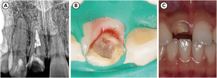
Figure 1 The initial periapical radiograph (A) and photography (B) shows that the fiber-reinforced composite post-resin core of upper maxillary (right) central incisor is partially broken.

Figure 2 Planning of coronal, sagittal, and axial plane of the guide drill placement for removal of fiber-reinforced composite post using the implant planning software (DDS-Pro).

Figure 3 (A) The stent is positioned in the mouth. (B) After 8.0 mm apical advancement of the guide drill. (C) After removing the remaining fiber-reinforced composite post-resin core under the dental operating microscope. (D) Radiographic verification of post removal. (E) After seating cast post and core and full veneer crown. (F) Radiography of completed case.
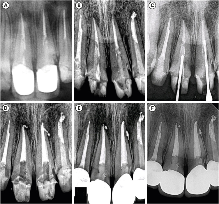
Figure 4 (A) Preoperative radiography shows preinstalled fiber-reinforced composite (FRC) posts and periapical lesions in the upper maxillary (right) central incisor, upper maxillary (left) central incisor, and upper maxillary (left) lateral incisor. (B) For the upper maxillary (left) lateral incisor, the FRC post was removed with a conventional method, and nonsurgical root canal treatment was completed. (C) For upper maxillary (right) and (left) central incisors, guided endodontics was used for removing the FRC posts. Periapical radiography shows the direction of the guide drill. (D) After removing the remaining FRC post with an ultrasonic tip, the root canal was obturated nonsurgically. (E) One-month follow-up radiograph. (F) The periapical lesion was resolved after 1-year follow-up.
Reference
-
1. Goodacre CJ, Spolnik KJ. The prosthodontic management of endodontically treated teeth: a literature review. Part I. Success and failure data, treatment concepts. J Prosthodont. 1994; 3:243–250. PMID: 7866508.
Article3. Cagidiaco MC, Goracci C, Garcia-Godoy F, Ferrari M. Clinical studies of fiber posts: a literature review. Int J Prosthodont. 2008; 21:328–336. PMID: 18717092.4. Lindemann M, Yaman P, Dennison JB, Herrero AA. Comparison of the efficiency and effectiveness of various techniques for removal of fiber posts. J Endod. 2005; 31:520–522. PMID: 15980712.
Article5. Gesi A, Magnolfi S, Goracci C, Ferrari M. Comparison of two techniques for removing fiber posts. J Endod. 2003; 29:580–582. PMID: 14503831.
Article6. Scotti N, Bergantin E, Alovisi M, Pasqualini D, Berutti E. Evaluation of a simplified fiber post removal system. J Endod. 2013; 39:1431–1434. PMID: 24139268.
Article7. Haupt F, Pfitzner J, Hülsmann M. A comparative in vitro study of different techniques for removal of fibre posts from root canals. Aust Endod J. 2018; 44:245–250. PMID: 28940721.
Article8. Krastl G, Zehnder MS, Connert T, Weiger R, Kühl S. Guided endodontics: a novel treatment approach for teeth with pulp canal calcification and apical pathology. Dent Traumatol. 2016; 32:240–246. PMID: 26449290.
Article9. van der Meer WJ, Vissink A, Ng YL, Gulabivala K 3rd. 3D computer aided treatment planning in endodontics. J Dent. 2016; 45:67–72. PMID: 26627596.
Article10. Lara-Mendes STO, Barbosa CFM, Santa-Rosa CC, Machado VC. Guided endodontic access in maxillary molars using cone-beam computed tomography and computer-aided design/computer-aided manufacturing system: a case report. J Endod. 2018; 44:875–879. PMID: 29571910.
Article11. Orstavik D, Kerekes K, Eriksen HM. The periapical index: a scoring system for radiographic assessment of apical periodontitis. Endod Dent Traumatol. 1986; 2:20–34. PMID: 3457698.
Article12. Hüfner T, Geerling J, Oldag G, Richter M, Kfuri M Jr, Pohlemann T, Krettek C. Accuracy study of computer-assisted drilling: the effect of bone density, drill bit characteristics, and use of a mechanical guide. J Orthop Trauma. 2005; 19:317–322. PMID: 15891540.13. Maia LM, Moreira Júnior G, Albuquerque RC, de Carvalho Machado V, da Silva NRFA, Hauss DD, da Silveira RR. Three-dimensional endodontic guide for adhesive fiber post removal: a dental technique. J Prosthet Dent. 2019; 121:387–390. PMID: 30477921.
Article14. Perez C, Finelle G, Couvrechel C. Optimisation of a guided endodontics protocol for removal of fibre-reinforced posts. Aust Endod J. 2020; 46:107–114. PMID: 31603599.
Article15. Zehnder MS, Connert T, Weiger R, Krastl G, Kühl S. Guided endodontics: accuracy of a novel method for guided access cavity preparation and root canal location. Int Endod J. 2016; 49:966–972. PMID: 26353942.
Article16. Connert T, Zehnder MS, Weiger R, Kühl S, Krastl G. Microguided endodontics: accuracy of a miniaturized technique for apically extended access cavity preparation in anterior teeth. J Endod. 2017; 43:787–790. PMID: 28292595.
Article
- Full Text Links
-
- Actions
-
Cited
- CITED
-
- Close
- Share
-
- Similar articles
-
- Currently there are so many fiber reinforced composite posts in the market. Some products are factory silanated but some products are not. Should I use silane for surface treatment of fiber reinforced composite posts?
- Fiber-reinforced composite resin bridges: an alternative method to treat root-fractured teeth
- Effect of fiber direction on the polymerization shrinkage of fiber-reinforced composites
- Reattachment of a fractured fragment with relined fiber post using indirect technique: a case report
- The influence of fitness and type of luting agents on bonding strength of fiber-reinforced composite resin posts
