Restor Dent Endod.
2021 Aug;46(3):e34. 10.5395/rde.2021.46.e34.
Evaluation of the relation between the pulp stones and direct restorations using cone beam computed tomography in a Turkish subpopulation
- Affiliations
-
- 1Department of Endodontics, Faculty of Dentistry, Biruni University, İstanbul, Turkey
- KMID: 2548078
- DOI: http://doi.org/10.5395/rde.2021.46.e34
Abstract
Objectives
This study aimed to assess the presence of pulp stones through an examination of cone beam computed tomography images and correlate their prevalence with age, sex, dental arch and side, tooth type, and restoration type and depth.
Materials and Methods
Cone beam computed tomography images obtained from 673 patients and archival data on 11,494 teeth were evaluated. The associations of pulp stones with age, sex, dental arch and side, tooth type, and restoration type and depth were noted. All the measurements were subjected to a χ2 test and one sample χ2 test (p < 0.05).
Results
In the study group, 163 (24.2%) patients and 379 (3.3%) teeth had at least one pulp stone. The pulp stone frequency in those aged 30–39 years was significantly greater than in those aged 18–29 and ≥ 60 years, and the frequency was higher in females than in males (p < 0.05). The highest prevalence of pulp stones was found in maxillary dental arches and molar teeth (p < 0.05). Pulp stones were significantly more common in medium-depth restorations (p < 0.05).
Conclusions
Maxillary molar teeth, medium-depth restorations, individuals aged 30–39 years and females had a greater percentage of pulp stones.
Figure
-
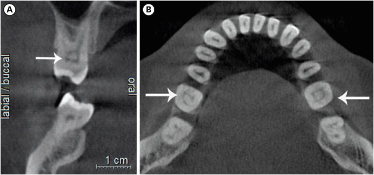
Figure 1 Teeth with pulp stones on different sections of cone-beam computed tomography (CBCT). (A) Coronal CBCT section of maxillary first molar. (B) Axial CBCT section of mandibular first molar.
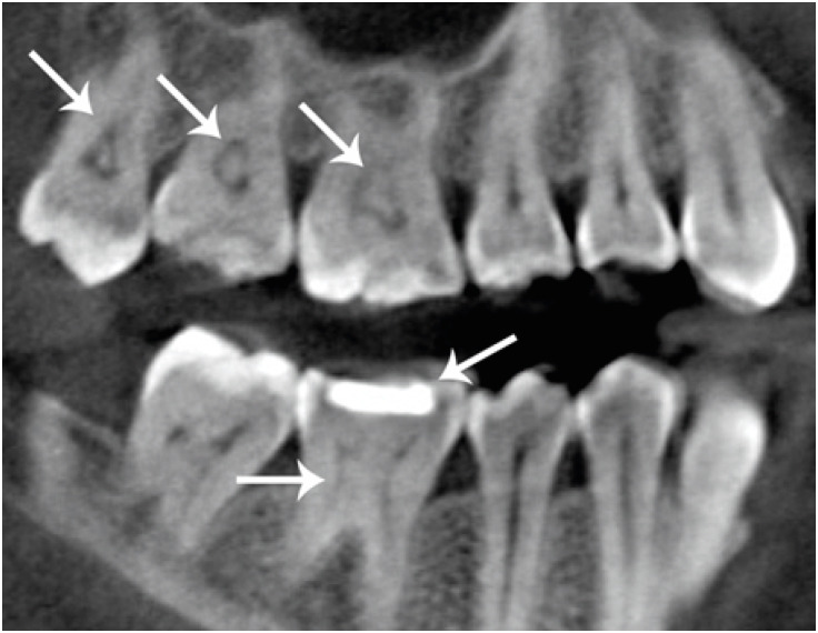
Figure 2 Sagittal cone-beam computed tomography section of maxillary molars with pulp stones and a mandibular first molar with a pulp stone and medium restoration.
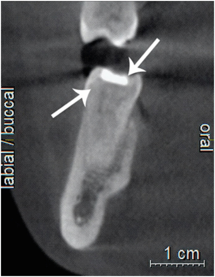
Figure 3 Coronal cone-beam computed tomography section of a mandibular first molar with a pulp stone and medium restoration.
Reference
-
1. Bevelander G, Johnson PL. Histogenesis and histochemistry of pulpal calcification. J Dent Res. 1956; 35:714–722. PMID: 13367289.
Article2. Goga R, Chandler NP, Oginni AO. Pulp stones: a review. Int Endod J. 2008; 41:457–468. PMID: 18422587.
Article3. Milcent CPF, da Silva TG, Baika LM, Grassi MT, Carneiro E, Franco A, de Lima AAS. Morphologic, structural, and chemical properties of pulp stones in exracted human teeth. J Endod. 2019; 45:1504–1512. PMID: 31757339.
Article4. Berès F, Isaac J, Mouton L, Rouzière S, Berdal A, Simon S, Dessombz A. Comparative physicochemical analysis of pulp stone and dentin. J Endod. 2016; 42:432–438. PMID: 26794341.
Article5. Edds AC, Walden JE, Scheetz JP, Goldsmith LJ, Drisko CL, Eleazer PD. Pilot study of correlation of pulp stones with cardiovascular disease. J Endod. 2005; 31:504–506. PMID: 15980708.
Article6. VanDenBerghe JM, Panther B, Gound TG. Pulp stones throughout the dentition of monozygotic twins: a case report. Oral Surg Oral Med Oral Pathol Oral Radiol Endod. 1999; 87:749–751. PMID: 10397671.7. Zeng JF, Zhang W, Jiang HW, Ling JQ. Isolation, cultivation and initial identification of Nanobacteria from dental pulp stone. Zhonghua Kou Qiang Yi Xue Za Zhi. 2006; 41:498–501. PMID: 17074192.8. Nayak M, Kumar J, Prasad LK. A radiographic correlation between systemic disorders and pulp stones. Indian J Dent Res. 2010; 21:369–373. PMID: 20930347.
Article9. da Silva EJNL, Prado MC, Queiroz PM, Nejaim Y, Brasil DM, Groppo FC, Haiter-Neto F. Assessing pulp stones by cone-beam computed tomography. Clin Oral Investig. 2017; 21:2327–2333.
Article10. Sener S, Cobankara FK, Akgünlü F. Calcifications of the pulp chamber: prevalence and implicated factors. Clin Oral Investig. 2009; 13:209–215.
Article11. Hsieh CY, Wu YC, Su CC, Chung MP, Huang RY, Ting PY, Lai CK, Chang KS, Tsai YC, Shieh YS. The prevalence and distribution of radiopaque, calcified pulp stones: a cone-beam computed tomography study in a northern Taiwanese population. J Dent Sci. 2018; 13:138–144. PMID: 30895109.
Article12. Chaini K, Georgopoulou MK. General pulp calcification: literature review and case report. Endod Pract Today. 2016; 10:69–75.13. Kannan S, Kannepady SK, Muthu K, Jeevan MB, Thapasum A. Radiographic assessment of the prevalence of pulp stones in Malaysians. J Endod. 2015; 41:333–337. PMID: 25476972.14. Turkal M, Tan E, Uzgur R, Hamidi M, Colak H, Uzgur Z. Incidence and distribution of pulp stones found in radiographic dental examination of adult Turkish dental patients. Ann Med Health Sci Res. 2013; 3:572–576. PMID: 24380011.
Article15. Patel S, Durack C, Abella F, Shemesh H, Roig M, Lemberg K. Cone beam computed tomography in Endodontics - a review. Int Endod J. 2015; 48:3–15. PMID: 24697513.16. Lari SS, Shokri A, Hosseinipanah SM, Rostami S, Sabounchi SS. Comparative sensitivity assessment of cone beam computed tomography and digital radiography for detecting foreign bodies. J Contemp Dent Pract. 2016; 17:224–229. PMID: 27207202.
Article17. Colak H, Celebi AA, Hamidi MM, Bayraktar Y, Colak T, Uzgur R. Assessment of the prevalence of pulp stones in a sample of Turkish Central Anatolian population. ScientificWorldJournal. 2012; 2012:804278. PMID: 22645455.18. Ranjitkar S, Taylor JA, Townsend GC. A radiographic assessment of the prevalence of pulp stones in Australians. Aust Dent J. 2002; 47:36–40. PMID: 12035956.
Article19. Verma KG, Juneja S, Randhawa S, Dhebar TM, Raheja A. Retrieval of iatrogenically pushed pulp stone from middle third of root canal in permanent maxillary central incisor: a case report. J Clin Diagn Res. 2015; 9:ZD06–ZD07.
Article20. Ibarrola JL, Knowles KI, Ludlow MO, McKinley IB Jr. Factors affecting the negotiability of second mesiobuccal canals in maxillary molars. J Endod. 1997; 23:236–238. PMID: 9594773.
Article21. Kayal RA. Distortion of digital panoramic radiographs used for implant site assessment. J Orthod Sci. 2016; 5:117–120. PMID: 27843885.
Article22. Moss-Salentijn L, Hendricks-Klyvert M. Calcified structures in human dental pulps. J Endod. 1988; 14:184–189. PMID: 3077408.
Article23. Tassoker M, Magat G, Sener S. A comparative study of cone-beam computed tomography and digital panoramic radiography for detecting pulp stones. Imaging Sci Dent. 2018; 48:201–212. PMID: 30276157.
Article24. Patil SR, Ghani HA, Almuhaiza M, Al-Zoubi IA, Anil KN, Misra N, Raghuram P. Prevalence of pulp stones in a Saudi Arabian subpopulation: a cone-beam computed tomography study. Saudi Endod J. 2018; 8:93–98.
Article25. Arys A, Philippart C, Dourov N. Microradiography and light microscopy of mineralization in the pulp of undemineralized human primary molars. J Oral Pathol Med. 1993; 22:49–53. PMID: 8445542.
Article26. Gulsahi A, Cebeci AI, Ozden S. A radiographic assessment of the prevalence of pulp stones in a group of Turkish dental patients. Int Endod J. 2009; 42:735–739. PMID: 19549152.
Article27. Tamse A, Kaffe I, Littner MM, Shani R. Statistical evaluation of radiologic survey of pulp stones. J Endod. 1982; 8:455–458. PMID: 6958783.
Article28. Alaçam T. 31. Geriatrik endodonti. In: Endodonti. Ankara: Özyurt Matbaacılık;2012. p. 1178.29. al-Hadi Hamasha A, Darwazeh A. Prevalence of pulp stones in Jordanian adults. Oral Surg Oral Med Oral Pathol Oral Radiol Endod. 1998; 86:730–732. PMID: 9868733.
Article
- Full Text Links
-
- Actions
-
Cited
- CITED
-
- Close
- Share
-
- Similar articles
-
- A comparative study of cone-beam computed tomography and digital panoramic radiography for detecting pulp stones
- Evaluation of morphometric features of fossa navicularis using cone-beam computed tomography in a Turkish subpopulation
- Management of root canal perforation by using cone-beam computed tomography
- Three-dimensional imaging modalities in endodontics
- A rare case of dilated invaginated odontome with talon cusp in a permanent maxillary central incisor diagnosed by cone beam computed tomography
