Color assessment of resin composite by using cellphone images compared with a spectrophotometer
- Affiliations
-
- 1Graduate Program in Dentistry, Federal University of Sergipe, Aracaju, SE, Brazil
- 2Graduate Program in Dentistry, Federal University of Pelotas, Pelotas, RS, Brazil
- 3Department of Dentistry, Federal University of Sergipe, Aracaju, SE, Brazil
- KMID: 2548067
- DOI: http://doi.org/10.5395/rde.2021.46.e23
Abstract
Objectives
This study assessed the reliability of digital color measurements using images of resin composite specimens captured with a cellphone.
Materials and Methods
The reference color of cylindrical specimens built-up with the use of resin composite (shades A1, A2, A3, and A4) was measured with a portable spectrophotometer (CIELab). Images of the specimens were obtained individually or pairwise (compared shades in the same photograph) under standardized parameters. The color of the specimens was measured in the images using RGB system and converted to CIELab system using image processing software. Whiteness index (WID ) and color differences (ΔE00 ) were calculated for each color measurement method. For the cellphone, the ΔE00 was calculated between the pairs of shades in separate images and in the same image. Data were analyzed using 2-way repeatedmeasures analysis of variance (α = 0.05). Linear regression models were used to predict the reference ΔE00 values of those calculated using color measured in the images.
Results
Images captured with the cellphone resulted in different WID values from the spectrophotometer only for shades A3 and A4. No difference to the reference ΔE00 was observed when individual images were used. In general, a similar ranking of ΔE00 among resin composite shades was observed for all methods. Stronger correlation coefficients with the reference ΔE00 were observed using individual than pairwise images.
Conclusions
This study showed that the use of cellphone images to measure the color difference seems to be a feasible alternative providing outcomes similar to those obtained with the spectrophotometer.
Figure
-
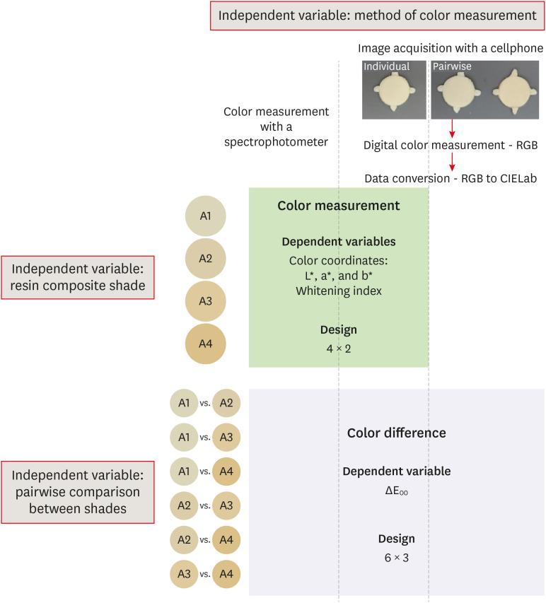
Figure 1 Schematic illustration of the experimental design of the study.
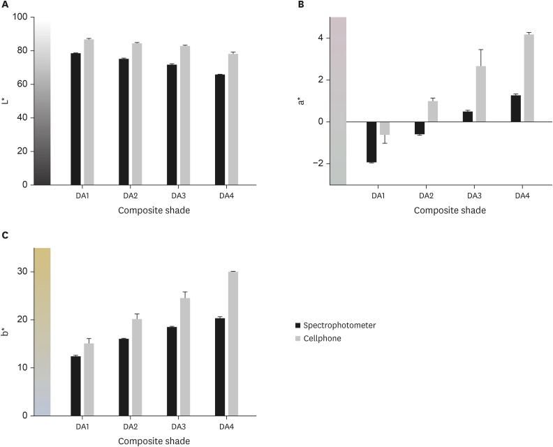
Figure 2 The behavior of color coordinates according to composite shade and method of color measurement. (A) Coordinate L* measuring lightness; (B) coordinate a* for red-to-green axis; and (C) coordinate b* for yellow-to-blue axis. Cellphone images presented the highest values for all coordinates.
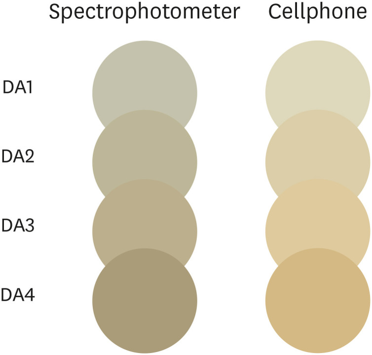
Figure 3 Illustrative cylinder-shaped composite specimens drawn using CorelDraw Graphics Suit X8 software and colored with Lab data obtained with each method of color measurement to simulate differences in color readings. In general, cellphone images appear lighter, redder, and yellower.

Figure 4 The 95% confidence interval (CI) for ∆E00 calculated using a spectrophotometer, or individual and pairwise images obtained from a cellphone (n = 9). CIs for all comparisons using individual images include the mean of the reference (spectrophotometer) of the respective comparison.

Figure 5 Scatterplots (left) and Linear regressions (right) were used to estimate the ∆E00 calculated using the spectrophotometer data as a function of values of ∆E00 calculated using images obtained with the cellphone. The predictive model improved as a coefficient of determination (R2) increased. (A, B) Individual images (R2 = 0.935); (C, D) pairwise images (R2 = 0.874). Coefficient (standard error).
Cited by 1 articles
-
Comparison of instrumental methods for color change assessment of Giomer resins
Luiza de Almeida Queiroz Ferreira, Rogéli Tibúrcio Ribeiro da Cunha Peixoto, Cláudia Silami de Magalhães, Tassiana Melo Sá, Monica Yamauti, Francisca Daniele Moreira Jardilino
Restor Dent Endod. 2022;47(1):e8. doi: 10.5395/rde.2022.47.e8.
Reference
-
1. Trifkovic B, Powers JM, Paravina RD. Color adjustment potential of resin composites. Clin Oral Investig. 2018; 22:1601–1607.
Article2. Westland S, Luo W, Li Y, Pan Q, Joiner A. Investigation of the perceptual thresholds of tooth whiteness. J Dent. 2017; 67S:S11–S14. PMID: 29233258.
Article3. Correa AC, Santana TR, Nahsan FP, Loguercio AD, Faria-E-Silva AL. The impact of a customized tray on in-office bleaching tooth sensitivity: a randomized clinical trial. Oper Dent. 2016; 41:15–22. PMID: 26244263.
Article4. Fernandes MT, Vaez SC, Lima CM, Nahsan FP, Loguércio AD, Faria-E-Silva AL. Preemptive use of naproxen on tooth sensitivity caused by in-office bleaching: a triple-blind, crossover, randomized clinical trial. Oper Dent. 2017; 42:486–496. PMID: 28829936.
Article5. Peixoto AC, Vaez SC, Soares KD, Ferreira LF, Loguercio AD, Faria-E-Silva AL. Preemptive use of piroxicam on tooth sensitivity caused by in-office bleaching: a randomized clinical trial. Braz Dent J. 2019; 30:498–504. PMID: 31664303.
Article6. Gokce HS, Piskin B, Ceyhan D, Gokce SM, Arisan V. Shade matching performance of normal and color vision-deficient dental professionals with standard daylight and tungsten illuminants. J Prosthet Dent. 2010; 103:139–147. PMID: 20188236.
Article7. Imbery TA, Tran D, Baechle MA, Hankle JL, Janus C. Dental shade matching and value discernment abilities of first-year dental students. J Prosthodont. 2018; 27:821–827. PMID: 29533495.
Article8. Corcodel N, Krisam J, Klotz AL, Deisenhofer UK, Stober T, Hassel AJ, Rammelsberg P, Zenthöfer A. Evaluation of small-group education on the shade determination ability of preclinical dental students-a controlled clinical trial. Eur J Dent Educ. 2018; 22:e582–e587. PMID: 29665167.
Article9. Rezende M, Chemin K, Vaez SC, Peixoto AC, Rabelo JF, Braga SSL, Faria-E-Silva AL, Silva GR, Soares CJ, Loguercio AD, Reis A. Effect of topical application of dipyrone on dental sensitivity reduction after in-office dental bleaching: a randomized, triple-blind multicenter clinical trial. J Am Dent Assoc. 2018; 149:363–371. PMID: 29550020.
Article10. Klaric Sever E, Budimir Z, Cerovac M, Stambuk M, Par M, Negovetic Vranic D, Tarle Z. Clinical and patient reported outcomes of bleaching effectiveness. Acta Odontol Scand. 2018; 76:30–38. PMID: 28893130.
Article11. Vaez SC, Correia A, Santana TR, Santana M, Peixoto AC, Leal PC, Faria-E-Silva AL. Is a single preliminary session of in-office bleaching beneficial for the effectiveness of at-home tooth bleaching? a randomized controlled clinical trial. Oper Dent. 2019; 44:E180–E189. PMID: 30849011.
Article12. Pereira R, Corado D, Silveira J, Alves R, Mata A, Marques D. Dental prophylaxis influence in tooth color assessment-clinical study. J Esthet Restor Dent. 2020; 32:586–592. PMID: 32400106.
Article13. Wee AG, Meyer A, Wu W, Wichman CS. Lighting conditions used during visual shade matching in private dental offices. J Prosthet Dent. 2016; 115:469–474. PMID: 26723088.
Article14. Igiel C, Lehmann KM, Ghinea R, Weyhrauch M, Hangx Y, Scheller H, Paravina RD. Reliability of visual and instrumental color matching. J Esthet Restor Dent. 2017; 29:303–308. PMID: 28742283.
Article15. Sullivan C, Pan Q, Westland S, Ellwood R. A yellowness index for use in dentistry. J Dent. 2019; 91:103244. PMID: 31730788.
Article16. Chu SJ, Trushkowsky RD, Paravina RD. Dental color matching instruments and systems. Review of clinical and research aspects. J Dent. 2010; 38(Supplement 2):e2–e16. PMID: 20621154.
Article17. Joiner A, Luo W. Tooth colour and whiteness: a review. J Dent. 2017; 67S:S3–S10. PMID: 28928097.
Article18. Gómez-Polo C, Portillo Muñoz M, Lorenzo Luengo MC, Vicente P, Galindo P, Martín Casado AM. Comparison of the CIELab and CIEDE2000 color difference formulas. J Prosthet Dent. 2016; 115:65–70. PMID: 26412001.
Article19. Pérez MM, Ghinea R, Rivas MJ, Yebra A, Ionescu AM, Paravina RD, Herrera LJ. Development of a customized whiteness index for dentistry based on CIELAB color space. Dent Mater. 2016; 32:461–467. PMID: 26778404.
Article20. Luo MR, Cui BR, Rigg B. The development of the CIE 2000 colour‐difference formula: CIEDE2000. Color Res Appl. 2001; 26:340–350.
Article21. Brook AH, Smith RN, Lath DJ. The clinical measurement of tooth colour and stain. Int Dent J. 2007; 57:324–330. PMID: 17992918.
Article22. Bhandari R, Thakur S, Singhal P, Chauhan D, Jayam C, Jain T. In vivo comparative evaluation of esthetics after microabrasion and microabrasion followed by casein phosphopeptide-amorphous calcium fluoride phosphate on molar incisor hypomineralization-affected incisors. Contemp Clin Dent. 2019; 10:9–15. PMID: 32015635.
Article23. Jarad FD, Russell MD, Moss BW. The use of digital imaging for colour matching and communication in restorative dentistry. Br Dent J. 2005; 199:43–49. PMID: 16003426.
Article24. Yamanel K, Caglar A, Özcan M, Gulsah K, Bagis B. Assessment of color parameters of composite resin shade guides using digital imaging versus colorimeter. J Esthet Restor Dent. 2010; 22:379–388. PMID: 21126293.
Article25. Tam WK, Lee HJ. Dental shade matching using a digital camera. J Dent. 2012; 40(Supplement 2):e3–e10.
Article26. Guan YH, Lath DL, Lilley TH, Willmot DR, Marlow I, Brook AH. The measurement of tooth whiteness by image analysis and spectrophotometry: a comparison. J Oral Rehabil. 2005; 32:7–15. PMID: 15634295.
Article27. Pérez MM, Herrera LJ, Carrillo F, Pecho OE, Dudea D, Gasparik C, Ghinea R, Bona AD. Whiteness difference thresholds in dentistry. Dent Mater. 2019; 35:292–297. PMID: 30527588.
Article28. Pecho OE, Ghinea R, do Amaral EA, Cardona JC, Della Bona A, Pérez MM. Relevant optical properties for direct restorative materials. Dent Mater. 2016; 32:e105–e112. PMID: 26994880.
Article29. Santos SMM, Silva PD, Faria-E-Silva AL. Color changes caused by reduction on the dentin shade composite thickness. Braz Dent J. 2018; 29:469–474. PMID: 30517446.
Article30. Dain SJ. Illuminant and observer metamerism and the Hardy-Rand-Rittler color vision test editions. Vis Neurosci. 2006; 23:685–694. PMID: 16998976.
Article31. Spink LS, Rungruanganut P, Megremis S, Kelly JR. Comparison of an absolute and surrogate measure of relative translucency in dental ceramics. Dent Mater. 2013; 29:702–707. PMID: 23618557.
Article32. Tung OH, Lai YL, Ho YC, Chou IC, Lee SY. Development of digital shade guides for color assessment using a digital camera with ring flashes. Clin Oral Investig. 2011; 15:49–56.
Article33. Lazar R, Culic B, Gasparik C, Lazar C, Dudea D. The accuracy of dental shade matching using cross-polarization photography. Int J Comput Dent. 2019; 22:343–351. PMID: 31840142.
- Full Text Links
-
- Actions
-
Cited
- CITED
-
- Close
- Share
-
- Similar articles
-
- Understanding of the color in composite resin
- Influence of the color of composite resins applied to lingual surface on the labial tooth color
- Color Stability of Alkasite Restorative Material: in vitro Studies
- Colorimetric comparison of single layered dental composite with double layered dental composite
- Evaluation of the Color Adjustment Potential of Single-Shade Composite Resin in Primary Teeth
